7TQO

Structure of human TREX1


Experimental Data Snapshot

  • Method: X-RAY DIFFRACTION
  • Resolution: 1.25 Å
  • R-Value Free: 0.190 
  • R-Value Work: 0.171 
  • R-Value Observed: 0.172 

wwPDB Validation   3D Report Full Report


This is version 1.1 of the entry. See complete history


Literature

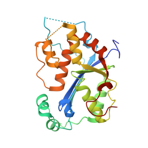
Structural basis of human TREX1 DNA degradation and autoimmune disease.

Zhou, W.Richmond-Buccola, D.Wang, Q.Kranzusch, P.J.

(2022) Nat Commun 13: 4277-4277

  • DOI: https://doi.org/10.1038/s41467-022-32055-z
  • Primary Citation of Related Structures:  
    7TQN, 7TQO, 7TQP, 7TQQ

  • PubMed Abstract: 

    TREX1 is a cytosolic DNA nuclease essential for regulation of cGAS-STING immune signaling. Existing structures of mouse TREX1 establish a mechanism of DNA degradation and provide a key model to explain autoimmune disease, but these structures incompletely explain human disease-associated mutations and have limited ability to guide development of small-molecule therapeutics. Here we determine crystal structures of human TREX1 in apo and DNA-bound conformations that provide high-resolution detail of all human-specific features. A 1.25 Å structure of human TREX1 establishes a complete model of solvation of the exonuclease active site and a 2.2 Å structure of the human TREX1-DNA complex enables identification of specific substitutions involved in DNA recognition. We map each TREX1 mutation associated with autoimmune disease and establish distinct categories of substitutions predicted to impact enzymatic function, protein stability, and interaction with cGAS-DNA liquid droplets. Our results explain how human-specific substitutions regulate TREX1 function and provide a foundation for structure-guided design of TREX1 therapeutics.


  • Organizational Affiliation

    Department of Immunology and Microbiology, School of Life Sciences, Southern University of Science and Technology, Shenzhen, Guangdong, 518055, China. zhouw@sustech.edu.cn.


Macromolecules
Find similar proteins by:  (by identity cutoff)  |  3D Structure
Entity ID: 1
MoleculeChains Sequence LengthOrganismDetailsImage
Three-prime repair exonuclease 1243Homo sapiensMutation(s): 8 
Gene Names: TREX1
EC: 3.1.11.2
UniProt & NIH Common Fund Data Resources
Find proteins for Q9NSU2 (Homo sapiens)
Explore Q9NSU2 
Go to UniProtKB:  Q9NSU2
PHAROS:  Q9NSU2
GTEx:  ENSG00000213689 
Entity Groups  
Sequence Clusters30% Identity50% Identity70% Identity90% Identity95% Identity100% Identity
UniProt GroupQ9NSU2
Sequence Annotations
Expand
  • Reference Sequence
Experimental Data & Validation

Experimental Data

  • Method: X-RAY DIFFRACTION
  • Resolution: 1.25 Å
  • R-Value Free: 0.190 
  • R-Value Work: 0.171 
  • R-Value Observed: 0.172 
  • Space Group: P 41 21 2
Unit Cell:
Length ( Å )Angle ( ˚ )
a = 57.592α = 90
b = 57.592β = 90
c = 124.4γ = 90
Software Package:
Software NamePurpose
PHENIXrefinement
XDSdata reduction
Aimlessdata scaling
PHASERphasing

Structure Validation

View Full Validation Report


Entry History & Funding Information

Deposition Data


Funding OrganizationLocationGrant Number
National Institutes of Health/National Institute of General Medical Sciences (NIH/NIGMS)United States1DP2GM146250-01

Revision History  (Full details and data files)

  • Version 1.0: 2022-08-03
    Type: Initial release
  • Version 1.1: 2023-10-18
    Changes: Data collection, Refinement description