7RM6

Horse liver alcohol dehydrogenase in complex with NADH and N-cylcohexyl formamide


Experimental Data Snapshot

  • Method: X-RAY DIFFRACTION
  • Resolution: 1.43 Å
  • R-Value Free: 0.208 
  • R-Value Work: 0.190 
  • R-Value Observed: 0.191 

wwPDB Validation   3D Report Full Report


Ligand Structure Quality Assessment 


This is version 1.2 of the entry. See complete history


Literature

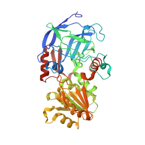
A two-directional vibrational probe reveals different electric field orientations in solution and an enzyme active site.

Zheng, C.Mao, Y.Kozuch, J.Atsango, A.O.Ji, Z.Markland, T.E.Boxer, S.G.

(2022) Nat Chem 14: 891-897

  • DOI: https://doi.org/10.1038/s41557-022-00937-w
  • Primary Citation of Related Structures:  
    7RM6

  • PubMed Abstract: 

    The catalytic power of an electric field depends on its magnitude and orientation with respect to the reactive chemical species. Understanding and designing new catalysts for electrostatic catalysis thus requires methods to measure the electric field orientation and magnitude at the molecular scale. We demonstrate that electric field orientations can be extracted using a two-directional vibrational probe by exploiting the vibrational Stark effect of both the C=O and C-D stretches of a deuterated aldehyde. Combining spectroscopy with molecular dynamics and electronic structure partitioning methods, we demonstrate that, despite distinct polarities, solvents act similarly in their preference for electrostatically stabilizing large bond dipoles at the expense of destabilizing small ones. In contrast, we find that for an active-site aldehyde inhibitor of liver alcohol dehydrogenase, the electric field orientation deviates markedly from that found in solvents, which provides direct evidence for the fundamental difference between the electrostatic environment of solvents and that of a preorganized enzyme active site.


  • Organizational Affiliation

    Department of Chemistry, Stanford University, Stanford, CA, USA.


Macromolecules
Find similar proteins by:  (by identity cutoff)  |  3D Structure
Entity ID: 1
MoleculeChains Sequence LengthOrganismDetailsImage
Alcohol dehydrogenase E chain
A, B
377Equus caballusMutation(s): 0 
EC: 1.1.1.1
UniProt
Find proteins for P00327 (Equus caballus)
Explore P00327 
Go to UniProtKB:  P00327
Entity Groups  
Sequence Clusters30% Identity50% Identity70% Identity90% Identity95% Identity100% Identity
UniProt GroupP00327
Sequence Annotations
Expand
  • Reference Sequence
Small Molecules
Ligands 3 Unique
IDChains Name / Formula / InChI Key2D Diagram3D Interactions
NAI
Query on NAI

Download Ideal Coordinates CCD File 
E [auth A],
I [auth B]
1,4-DIHYDRONICOTINAMIDE ADENINE DINUCLEOTIDE
C21 H29 N7 O14 P2
BOPGDPNILDQYTO-NNYOXOHSSA-N
CXF (Subject of Investigation/LOI)
Query on CXF

Download Ideal Coordinates CCD File 
F [auth A],
J [auth B]
CYCLOHEXYLFORMAMIDE
C7 H13 N O
SWGXDLRCJNEEGZ-UHFFFAOYSA-N
ZN
Query on ZN

Download Ideal Coordinates CCD File 
C [auth A],
D [auth A],
G [auth B],
H [auth B]
ZINC ION
Zn
PTFCDOFLOPIGGS-UHFFFAOYSA-N
Experimental Data & Validation

Experimental Data

  • Method: X-RAY DIFFRACTION
  • Resolution: 1.43 Å
  • R-Value Free: 0.208 
  • R-Value Work: 0.190 
  • R-Value Observed: 0.191 
  • Space Group: P 1
Unit Cell:
Length ( Å )Angle ( ˚ )
a = 44.142α = 92.41
b = 50.979β = 103.03
c = 92.864γ = 108.94
Software Package:
Software NamePurpose
PHENIXrefinement
Aimlessdata scaling
PDB_EXTRACTdata extraction
XDSdata reduction
PHENIXphasing

Structure Validation

View Full Validation Report


Ligand Structure Quality Assessment 


Entry History & Funding Information

Deposition Data


Funding OrganizationLocationGrant Number
National Institutes of Health/National Institute of General Medical Sciences (NIH/NIGMS)United StatesGM118044

Revision History  (Full details and data files)

  • Version 1.0: 2022-04-27
    Type: Initial release
  • Version 1.1: 2022-09-21
    Changes: Database references
  • Version 1.2: 2023-10-18
    Changes: Data collection, Refinement description