7QFE

Crystal structure of Human Serum albumin in complex with Gemfibrozil


Experimental Data Snapshot

  • Method: X-RAY DIFFRACTION
  • Resolution: 2.20 Å
  • R-Value Free: 0.255 
  • R-Value Work: 0.219 
  • R-Value Observed: 0.223 

wwPDB Validation   3D Report Full Report


Ligand Structure Quality Assessment 


This is version 1.1 of the entry. See complete history


Literature

Structural Analysis of Human Serum Albumin in Complex with the Fibrate Drug Gemfibrozil.

Liberi, S.Linciano, S.Moro, G.De Toni, L.Cendron, L.Angelini, A.

(2022) Int J Mol Sci 23

  • DOI: https://doi.org/10.3390/ijms23031769
  • Primary Citation of Related Structures:  
    7QFE

  • PubMed Abstract: 

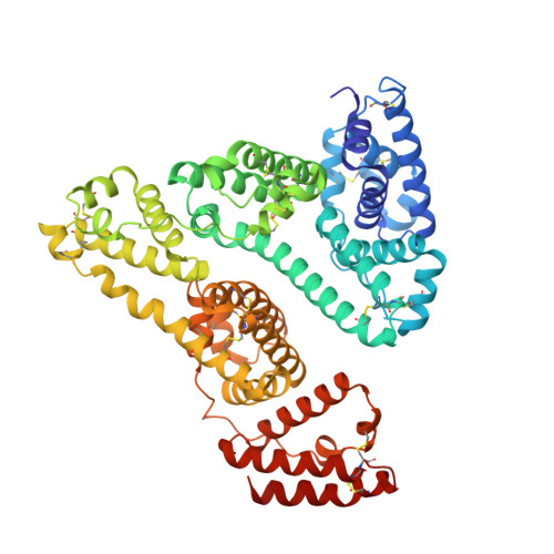
    Gemfibrozil (GEM) is an orally administered lipid-regulating fibrate derivative drug sold under the brand name Lopid ® , among others. Since its approval in the early 80s, GEM has been largely applied to treat hypertriglyceridemia and other disorders of lipid metabolism. Though generally well tolerated, GEM can alter the distribution and the free, active concentration of some co-administered drugs, leading to adverse effects. Most of them appear to be related to the ability of GEM to bind with high affinity human serum albumin (HSA), the major drug-carrier protein in blood plasma. Here, we report the crystal structure of HSA in complex with GEM. Two binding sites have been identified, namely Sudlow's binding sites I (FA7) and II (FA3-FA4). A comparison of the crystal structure of HSA in complex with GEM with those of other previously described HSA-drug complexes enabled us to appreciate the analogies and differences in their respective binding modes. The elucidation of the molecular interaction between GEM and HSA might offer the basis for the development of novel GEM derivatives that can be safely and synergistically co-administered with other drugs, enabling augmented therapeutic efficacies.


  • Organizational Affiliation

    Department of Biology, University of Padua, Viale G. Colombo 3, 35131 Padua, Italy.


Macromolecules
Find similar proteins by:  (by identity cutoff)  |  3D Structure
Entity ID: 1
MoleculeChains Sequence LengthOrganismDetailsImage
Serum albumin585Homo sapiensMutation(s): 0 
Gene Names: ALBGIG20GIG42PRO0903PRO1708PRO2044PRO2619PRO2675UNQ696/PRO1341
UniProt & NIH Common Fund Data Resources
Find proteins for P02768 (Homo sapiens)
Explore P02768 
Go to UniProtKB:  P02768
PHAROS:  P02768
GTEx:  ENSG00000163631 
Entity Groups  
Sequence Clusters30% Identity50% Identity70% Identity90% Identity95% Identity100% Identity
UniProt GroupP02768
Sequence Annotations
Expand
  • Reference Sequence
Small Molecules
Ligands 6 Unique
IDChains Name / Formula / InChI Key2D Diagram3D Interactions
4TX (Subject of Investigation/LOI)
Query on 4TX

Download Ideal Coordinates CCD File 
D [auth A],
M [auth A]
5-(2,5-dimethylphenoxy)-2,2-dimethylpentanoic acid
C15 H22 O3
HEMJJKBWTPKOJG-UHFFFAOYSA-N
MYR
Query on MYR

Download Ideal Coordinates CCD File 
B [auth A],
C [auth A]
MYRISTIC ACID
C14 H28 O2
TUNFSRHWOTWDNC-UHFFFAOYSA-N
PG4
Query on PG4

Download Ideal Coordinates CCD File 
F [auth A]TETRAETHYLENE GLYCOL
C8 H18 O5
UWHCKJMYHZGTIT-UHFFFAOYSA-N
PGE
Query on PGE

Download Ideal Coordinates CCD File 
E [auth A]TRIETHYLENE GLYCOL
C6 H14 O4
ZIBGPFATKBEMQZ-UHFFFAOYSA-N
MPD
Query on MPD

Download Ideal Coordinates CCD File 
G [auth A]
H [auth A]
I [auth A]
J [auth A]
K [auth A]
G [auth A],
H [auth A],
I [auth A],
J [auth A],
K [auth A],
L [auth A]
(4S)-2-METHYL-2,4-PENTANEDIOL
C6 H14 O2
SVTBMSDMJJWYQN-YFKPBYRVSA-N
PO4
Query on PO4

Download Ideal Coordinates CCD File 
N [auth A]PHOSPHATE ION
O4 P
NBIIXXVUZAFLBC-UHFFFAOYSA-K
Experimental Data & Validation

Experimental Data

  • Method: X-RAY DIFFRACTION
  • Resolution: 2.20 Å
  • R-Value Free: 0.255 
  • R-Value Work: 0.219 
  • R-Value Observed: 0.223 
  • Space Group: C 1 2 1
Unit Cell:
Length ( Å )Angle ( ˚ )
a = 184.897α = 90
b = 38.642β = 104.58
c = 96.213γ = 90
Software Package:
Software NamePurpose
REFMACrefinement
PDB_EXTRACTdata extraction
XDSdata reduction
STARANISOdata scaling
MOLREPphasing

Structure Validation

View Full Validation Report


Ligand Structure Quality Assessment 


Entry History & Funding Information

Deposition Data


Funding OrganizationLocationGrant Number
Other government--

Revision History  (Full details and data files)

  • Version 1.0: 2022-03-02
    Type: Initial release
  • Version 1.1: 2024-01-31
    Changes: Data collection, Refinement description