7P9O

Structure of E.coli RlmJ in complex with a SAM analogue (CA)


Experimental Data Snapshot

  • Method: X-RAY DIFFRACTION
  • Resolution: 2.10 Å
  • R-Value Free: 0.306 
  • R-Value Work: 0.240 
  • R-Value Observed: 0.243 

wwPDB Validation   3D Report Full Report


Ligand Structure Quality Assessment 


This is version 1.2 of the entry. See complete history


Literature

Synthesis of RNA-cofactor conjugates and structural exploration of RNA recognition by an m6A RNA methyltransferase.

Meynier, V.Iannazzo, L.Catala, M.Oerum, S.Braud, E.Atdjian, C.Barraud, P.Fonvielle, M.Tisne, C.Etheve-Quelquejeu, M.

(2022) Nucleic Acids Res 50: 5793-5806

  • DOI: https://doi.org/10.1093/nar/gkac354
  • Primary Citation of Related Structures:  
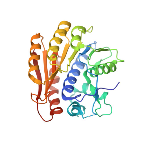
    7P8Q, 7P9I, 7P9O

  • PubMed Abstract: 

    Chemical synthesis of RNA conjugates has opened new strategies to study enzymatic mechanisms in RNA biology. To gain insights into poorly understood RNA nucleotide methylation processes, we developed a new method to synthesize RNA-conjugates for the study of RNA recognition and methyl-transfer mechanisms of SAM-dependent m6A RNA methyltransferases. These RNA conjugates contain a SAM cofactor analogue connected at the N6-atom of an adenosine within dinucleotides, a trinucleotide or a 13mer RNA. Our chemical route is chemo- and regio-selective and allows flexible modification of the RNA length and sequence. These compounds were used in crystallization assays with RlmJ, a bacterial m6A rRNA methyltransferase. Two crystal structures of RlmJ in complex with RNA-SAM conjugates were solved and revealed the RNA-specific recognition elements used by RlmJ to clamp the RNA substrate in its active site. From these structures, a model of a trinucleotide bound in the RlmJ active site could be built and validated by methyltransferase assays on RlmJ mutants. The methyl transfer by RlmJ could also be deduced. This study therefore shows that RNA-cofactor conjugates are potent molecular tools to explore the active site of RNA modification enzymes.


  • Organizational Affiliation

    Expression Génétique Microbienne, UMR 8261, CNRS, Université Paris Cité, Institut de Biologie Physico-Chimique (IBPC), 75005, Paris, France.


Macromolecules
Find similar proteins by:  (by identity cutoff)  |  3D Structure
Entity ID: 1
MoleculeChains Sequence LengthOrganismDetailsImage
Ribosomal RNA large subunit methyltransferase J280Escherichia coli K-12Mutation(s): 0 
Gene Names: rlmJyhiRb3499JW3466
EC: 2.1.1.266
UniProt
Find proteins for P37634 (Escherichia coli (strain K12))
Explore P37634 
Go to UniProtKB:  P37634
Entity Groups  
Sequence Clusters30% Identity50% Identity70% Identity90% Identity95% Identity100% Identity
UniProt GroupP37634
Sequence Annotations
Expand
  • Reference Sequence
Small Molecules
Ligands 1 Unique
IDChains Name / Formula / InChI Key2D Diagram3D Interactions
0Y0 (Subject of Investigation/LOI)
Query on 0Y0

Download Ideal Coordinates CCD File 
B [auth A]5'-{[(3S)-3-amino-3-carboxypropyl]amino}-5'-deoxyadenosine
C14 H21 N7 O5
WSOGVCAFRBSBRE-WFMPWKQPSA-N
Experimental Data & Validation

Experimental Data

  • Method: X-RAY DIFFRACTION
  • Resolution: 2.10 Å
  • R-Value Free: 0.306 
  • R-Value Work: 0.240 
  • R-Value Observed: 0.243 
  • Space Group: C 1 2 1
Unit Cell:
Length ( Å )Angle ( ˚ )
a = 79.99α = 90
b = 38.89β = 105.62
c = 90.65γ = 90
Software Package:
Software NamePurpose
XDSdata reduction
Aimlessdata scaling
MOLREPphasing
PHENIXrefinement
PDB_EXTRACTdata extraction

Structure Validation

View Full Validation Report


Ligand Structure Quality Assessment 


Entry History & Funding Information

Deposition Data


Funding OrganizationLocationGrant Number
Agence Nationale de la Recherche (ANR)FranceANR-19-CE07-0028

Revision History  (Full details and data files)

  • Version 1.0: 2022-05-25
    Type: Initial release
  • Version 1.1: 2022-06-15
    Changes: Database references
  • Version 1.2: 2024-01-31
    Changes: Data collection, Refinement description