7OVT

major seeded in vitro fibril morphology from murine SAA1.1 protein


Experimental Data Snapshot

  • Method: ELECTRON MICROSCOPY
  • Resolution: 2.69 Å
  • Aggregation State: HELICAL ARRAY 
  • Reconstruction Method: HELICAL 

wwPDB Validation   3D Report Full Report


This is version 1.0 of the entry. See complete history


Literature

Cryo-EM demonstrates the in vitro proliferation of an ex vivo amyloid fibril morphology by seeding.

Heerde, T.Rennegarbe, M.Biedermann, A.Savran, D.Pfeiffer, P.B.Hitzenberger, M.Baur, J.Puscalau-Girtu, I.Zacharias, M.Schwierz, N.Haupt, C.Schmidt, M.Fandrich, M.

(2022) Nat Commun 13: 85-85

  • DOI: https://doi.org/10.1038/s41467-021-27688-5
  • Primary Citation of Related Structures:  
    7OVT

  • PubMed Abstract: 

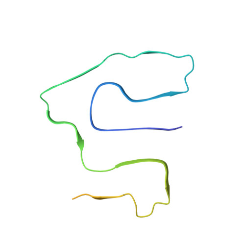
    Several studies showed that seeding of solutions of monomeric fibril proteins with ex vivo amyloid fibrils accelerated the kinetics of fibril formation in vitro but did not necessarily replicate the seed structure. In this research we use cryo-electron microscopy and other methods to analyze the ability of serum amyloid A (SAA)1.1-derived amyloid fibrils, purified from systemic AA amyloidosis tissue, to seed solutions of recombinant SAA1.1 protein. We show that 98% of the seeded fibrils remodel the full fibril structure of the main ex vivo fibril morphology, which we used for seeding, while they are notably different from unseeded in vitro fibrils. The seeded fibrils show a similar proteinase K resistance as ex vivo fibrils and are substantially more stable to proteolytic digestion than unseeded in vitro fibrils. Our data support the view that the fibril morphology contributes to determining proteolytic stability and that pathogenic amyloid fibrils arise from proteolytic selection.


  • Organizational Affiliation

    Institute of Protein Biochemistry, Ulm University, 89081, Ulm, Germany.


Macromolecules
Find similar proteins by:  (by identity cutoff)  |  3D Structure
Entity ID: 1
MoleculeChains Sequence LengthOrganismDetailsImage
Serum amyloid A-2 protein
A, B, C, D, E
A, B, C, D, E, F, G, H, I, J, K, L
103Mus musculusMutation(s): 0 
Gene Names: Saa2
UniProt
Find proteins for P05367 (Mus musculus)
Explore P05367 
Go to UniProtKB:  P05367
Entity Groups  
Sequence Clusters30% Identity50% Identity70% Identity90% Identity95% Identity100% Identity
UniProt GroupP05367
Sequence Annotations
Expand
  • Reference Sequence
Experimental Data & Validation

Experimental Data

  • Method: ELECTRON MICROSCOPY
  • Resolution: 2.69 Å
  • Aggregation State: HELICAL ARRAY 
  • Reconstruction Method: HELICAL 
EM Software:
TaskSoftware PackageVersion
MODEL REFINEMENTPHENIX1.16-3549
RECONSTRUCTIONRELION3.0.4

Structure Validation

View Full Validation Report


Entry History & Funding Information

Deposition Data


Funding OrganizationLocationGrant Number
German Research Foundation (DFG)GermanyDFG SCHM 3276/1
German Research Foundation (DFG)GermanyDFG FA 456/23
German Research Foundation (DFG)GermanyDFG FA 456/27
German Research Foundation (DFG)GermanyDFG HA 7138/3
German Research Foundation (DFG)GermanyDFG HA 7138/2
German Research Foundation (DFG)GermanyCRC 1279 project A03

Revision History  (Full details and data files)

  • Version 1.0: 2022-02-02
    Type: Initial release