7JQ8

Solution NMR structure of human Brd3 ET domain


Experimental Data Snapshot

  • Method: SOLUTION NMR
  • Conformers Calculated: 200 
  • Conformers Submitted: 20 
  • Selection Criteria: structures with the lowest energy 

wwPDB Validation   3D Report Full Report


This is version 1.2 of the entry. See complete history


Literature

A common binding motif in the ET domain of BRD3 forms polymorphic structural interfaces with host and viral proteins.

Aiyer, S.Swapna, G.V.T.Ma, L.C.Liu, G.Hao, J.Chalmers, G.Jacobs, B.C.Montelione, G.T.Roth, M.J.

(2021) Structure 29: 886

  • DOI: https://doi.org/10.1016/j.str.2021.01.010
  • Primary Citation of Related Structures:  
    7JMY, 7JQ8, 7JYN, 7JYZ

  • PubMed Abstract: 

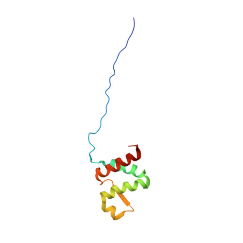
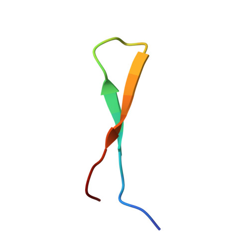
    The extraterminal (ET) domain of BRD3 is conserved among BET proteins (BRD2, BRD3, BRD4), interacting with multiple host and viral protein-protein networks. Solution NMR structures of complexes formed between the BRD3 ET domain and either the 79-residue murine leukemia virus integrase (IN) C-terminal domain (IN 329-408 ) or its 22-residue IN tail peptide (IN 386-407 ) alone reveal similar intermolecular three-stranded β-sheet formations. 15 N relaxation studies reveal a 10-residue linker region (IN 379-388 ) tethering the SH3 domain (IN 329-378 ) to the ET-binding motif (IN 389-405 ):ET complex. This linker has restricted flexibility, affecting its potential range of orientations in the IN:nucleosome complex. The complex of the ET-binding peptide of the host NSD3 protein (NSD3 148-184 ) and the BRD3 ET domain includes a similar three-stranded β-sheet interaction, but the orientation of the β hairpin is flipped compared with the two IN:ET complexes. These studies expand our understanding of molecular recognition polymorphism in complexes of ET-binding motifs with viral and host proteins.


  • Organizational Affiliation

    Department of Pharmacology, Robert Wood Johnson Medical School, Rutgers University, Piscataway, NJ 08854, USA; Department of Biochemistry and Molecular Biology, Robert Wood Johnson Medical School, Rutgers University, Piscataway, NJ 08854, USA.


Macromolecules
Find similar proteins by:  (by identity cutoff)  |  3D Structure
Entity ID: 1
MoleculeChains Sequence LengthOrganismDetailsImage
Bromodomain-containing protein 396Homo sapiensMutation(s): 0 
Gene Names: BRD3KIAA0043RING3L
UniProt & NIH Common Fund Data Resources
Find proteins for Q15059 (Homo sapiens)
Explore Q15059 
Go to UniProtKB:  Q15059
PHAROS:  Q15059
GTEx:  ENSG00000169925 
Entity Groups  
Sequence Clusters30% Identity50% Identity70% Identity90% Identity95% Identity100% Identity
UniProt GroupQ15059
Sequence Annotations
Expand
  • Reference Sequence

Find similar proteins by:  Sequence   |   3D Structure  

Entity ID: 2
MoleculeChains Sequence LengthOrganismDetailsImage
Integrase peptide23Moloney murine leukemia virus isolate ShinnickMutation(s): 0 
Gene Names: gag-pol
EC: 2.7.7 (PDB Primary Data), 3.1 (PDB Primary Data)
UniProt
Find proteins for P03355 (Moloney murine leukemia virus (isolate Shinnick))
Explore P03355 
Go to UniProtKB:  P03355
Entity Groups  
Sequence Clusters30% Identity50% Identity70% Identity90% Identity95% Identity100% Identity
UniProt GroupP03355
Sequence Annotations
Expand
  • Reference Sequence
Experimental Data & Validation

Experimental Data

  • Method: SOLUTION NMR
  • Conformers Calculated: 200 
  • Conformers Submitted: 20 
  • Selection Criteria: structures with the lowest energy 

Structure Validation

View Full Validation Report


Entry History & Funding Information

Deposition Data


Funding OrganizationLocationGrant Number
National Institutes of Health/Office of the DirectorUnited StatesNIH-MIRA R35GM122518
National Institutes of Health/Office of the DirectorUnited StatesRO1 GM110639
National Institutes of Health/Office of the DirectorUnited StatesGM120574

Revision History  (Full details and data files)

  • Version 1.0: 2021-06-23
    Type: Initial release
  • Version 1.1: 2021-08-18
    Changes: Database references
  • Version 1.2: 2023-06-14
    Changes: Other