7CD1

Crystal structure of inhibitory Smad, Smad7


Experimental Data Snapshot

  • Method: X-RAY DIFFRACTION
  • Resolution: 1.89 Å
  • R-Value Free: 0.199 
  • R-Value Work: 0.174 
  • R-Value Observed: 0.175 

wwPDB Validation   3D Report Full Report


This is version 1.1 of the entry. See complete history


Literature

Structural basis for inhibitory effects of Smad7 on TGF-beta family signaling.

Murayama, K.Kato-Murayama, M.Itoh, Y.Miyazono, K.Miyazawa, K.Shirouzu, M.

(2020) J Struct Biol 212: 107661-107661

  • DOI: https://doi.org/10.1016/j.jsb.2020.107661
  • Primary Citation of Related Structures:  
    7CD1

  • PubMed Abstract: 

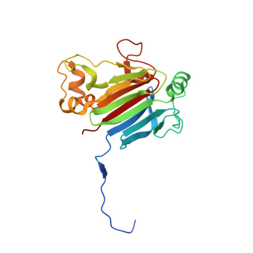
    Smad6 and Smad7 are classified as inhibitory Smads (I-Smads). They are crucial in the fine-tuning of signals by cytokines of the transforming growth factor-β (TGF-β) family. They are negative feedback regulators and principally target the activated type I receptors as well as the activated Smad complexes, but with distinct specificities. Smad7 inhibits Smad signaling from all seven type I receptors of the TGF-β family, whereas Smad6 preferentially inhibits Smad signaling from the bone morphogenetic protein (BMP) type I receptors, BMPR1A and BMPR1B. The target specificities are attributed to the C-terminal MH2 domain. Notably, Smad7 utilizes two alternative molecular surfaces for its inhibitory function against type I receptors. One is a basic groove composed of the first α-helix and the L3 loop, a structure that is shared with Smad6 and receptor-regulated Smads (R-Smads). The other is a three-finger-like structure (consisting of residues 331-361, 379-387, and the L3 loop) that is unique to Smad7. The underlying structural basis remains to be elucidated in detail. Here, we report the crystal structure of the MH2 domain of mouse Smad7 at 1.9 Å resolution. The three-finger-like structure is stabilized by a network of hydrogen bonds between residues 331-361 and 379-387, thus forming a molecular surface unique to Smad7. Furthermore, we discuss how Smad7 antagonizes the activated Smad complexes composed of R-Smad and Smad4, a common partner Smad.


  • Organizational Affiliation

    Laboratory for Protein Functional and Structural Biology, RIKEN Center for Biosystems Dynamics Research, Yokohama, 1-7-22 Suehiro-cho, Tsurumi, Yokohama 230-0045, Japan; Graduate School of Biomedical Engineering, Tohoku University, 2-1 Seiryomachi, Aoba, Sendai 980-8575, Japan.


Macromolecules
Find similar proteins by:  (by identity cutoff)  |  3D Structure
Entity ID: 1
MoleculeChains Sequence LengthOrganismDetailsImage
Mothers against decapentaplegic homolog 7
A, B, C, D
187Mus musculusMutation(s): 0 
Gene Names: Smad7Madh7Madh8
UniProt
Find proteins for O35253 (Mus musculus)
Explore O35253 
Go to UniProtKB:  O35253
Entity Groups  
Sequence Clusters30% Identity50% Identity70% Identity90% Identity95% Identity100% Identity
UniProt GroupO35253
Sequence Annotations
Expand
  • Reference Sequence
Small Molecules
Ligands 2 Unique
IDChains Name / Formula / InChI Key2D Diagram3D Interactions
SO4
Query on SO4

Download Ideal Coordinates CCD File 
E [auth A]
I [auth B]
J [auth B]
K [auth C]
L [auth C]
E [auth A],
I [auth B],
J [auth B],
K [auth C],
L [auth C],
M [auth C],
N [auth C],
Q [auth D]
SULFATE ION
O4 S
QAOWNCQODCNURD-UHFFFAOYSA-L
CL
Query on CL

Download Ideal Coordinates CCD File 
F [auth A]
G [auth A]
H [auth A]
O [auth C]
P [auth C]
F [auth A],
G [auth A],
H [auth A],
O [auth C],
P [auth C],
R [auth D],
S [auth D]
CHLORIDE ION
Cl
VEXZGXHMUGYJMC-UHFFFAOYSA-M
Experimental Data & Validation

Experimental Data

  • Method: X-RAY DIFFRACTION
  • Resolution: 1.89 Å
  • R-Value Free: 0.199 
  • R-Value Work: 0.174 
  • R-Value Observed: 0.175 
  • Space Group: C 2 2 21
Unit Cell:
Length ( Å )Angle ( ˚ )
a = 93.205α = 90
b = 117.426β = 90
c = 163.239γ = 90
Software Package:
Software NamePurpose
PHENIXrefinement
HKL-2000data reduction
HKL-2000data scaling
MOLREPphasing

Structure Validation

View Full Validation Report


Entry History 

Deposition Data

Revision History  (Full details and data files)

  • Version 1.0: 2021-03-17
    Type: Initial release
  • Version 1.1: 2023-11-29
    Changes: Data collection, Database references, Refinement description