7C5E

Crystal structure of Keap1 in complex with fumarate (FUM)


Experimental Data Snapshot

  • Method: X-RAY DIFFRACTION
  • Resolution: 1.75 Å
  • R-Value Free: 0.245 
  • R-Value Work: 0.202 
  • R-Value Observed: 0.204 

wwPDB Validation   3D Report Full Report


Ligand Structure Quality Assessment 


This is version 2.1 of the entry. See complete history


Literature

Structural insights into the multiple binding modes of Dimethyl Fumarate (DMF) and its analogs to the Kelch domain of Keap1.

Unni, S.Deshmukh, P.Krishnappa, G.Kommu, P.Padmanabhan, B.

(2021) FEBS J 288: 1599-1613

  • DOI: https://doi.org/10.1111/febs.15485
  • Primary Citation of Related Structures:  
    6LRZ, 7C5E, 7C60

  • PubMed Abstract: 

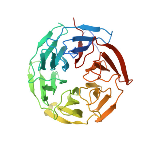
    The activation of the nuclear factor erythroid 2-related factor 2 (Nrf2) transcription function has been implicated in the protection of neurodegenerative diseases. The cytoplasmic protein, Kelch-like ECH-associated protein 1 (Keap1), negatively regulates Nrf2. The Keap1-Nrf2 pathway is a potential therapeutic target for tackling free-radical damage. Dimethyl fumarate (DMF) is currently an approved drug for the treatment of relapsing multiple sclerosis. Recent studies showed that DMF modifies the reactive cysteines in the BTB domain of Keap1 and thus activates Nrf2 transcription function. Intriguingly, our crystal structure studies revealed that DMF also binds to the β-propeller domain (Keap1-DC) of Keap1. The crystal structure of the complex, refined to 1.54 Å resolution, revealed unexpected features: DMF binds (a) to the Nrf2-binding site (bottom region of Keap1-DC, site 1) with moderate interaction, and (b) to the top region of Keap1-DC, near to the blade II (site 2). The specificity of the binding 'site 2' was found to be unique to blade II of the β-propeller domain. The newly identified 'site 2' region in Keap1-DC may have a different functional role to regulate Nrf2. Moreover, the crystal structures of Keap1-DC in complex with the DMF analogs, including monoethyl fumarate, fumarate, and itaconate, also exhibited similar binding modes with Keap1-DC. Binding studies confirmed that DMF binds, in a nanomolar range, to the Keap1-DC region as well as the BTB domain of Keap1. Furthermore, the competitive binding assay in the presence of the Nrf2 peptide affirmed the direct binding of DMF at the Nrf2-binding region of Keap1-DC. Overall, our studies suggest that the drug molecule, DMF, binds at multiple sites of Keap1 and thus potentially activates Nrf2 function through covalent as well as the noncovalent mode of action, to combat oxidative stress. DATABASE: Structural data are available in RCSB-protein data bank database(s) under the accession numbers 6LRZ, 7C60, and 7C5E.


  • Organizational Affiliation

    Department of Biophysics, National Institute of Mental Health and Neuro Sciences (NIMHANS), Bengaluru, India.


Macromolecules
Find similar proteins by:  (by identity cutoff)  |  3D Structure
Entity ID: 1
MoleculeChains Sequence LengthOrganismDetailsImage
Kelch-like ECH-associated protein 1314Mus musculusMutation(s): 0 
Gene Names: Keap1Inrf2Kiaa0132
UniProt
Find proteins for Q9Z2X8 (Mus musculus)
Explore Q9Z2X8 
Go to UniProtKB:  Q9Z2X8
Entity Groups  
Sequence Clusters30% Identity50% Identity70% Identity90% Identity95% Identity100% Identity
UniProt GroupQ9Z2X8
Sequence Annotations
Expand
  • Reference Sequence
Small Molecules
Ligands 4 Unique
IDChains Name / Formula / InChI Key2D Diagram3D Interactions
PGE
Query on PGE

Download Ideal Coordinates CCD File 
E [auth A]TRIETHYLENE GLYCOL
C6 H14 O4
ZIBGPFATKBEMQZ-UHFFFAOYSA-N
FUM (Subject of Investigation/LOI)
Query on FUM

Download Ideal Coordinates CCD File 
B [auth A],
C [auth A],
F [auth A]
FUMARIC ACID
C4 H4 O4
VZCYOOQTPOCHFL-OWOJBTEDSA-N
SO4
Query on SO4

Download Ideal Coordinates CCD File 
H [auth A],
I [auth A]
SULFATE ION
O4 S
QAOWNCQODCNURD-UHFFFAOYSA-L
ACT
Query on ACT

Download Ideal Coordinates CCD File 
D [auth A],
G [auth A]
ACETATE ION
C2 H3 O2
QTBSBXVTEAMEQO-UHFFFAOYSA-M
Binding Affinity Annotations 
IDSourceBinding Affinity
FUM Binding MOAD:  7C5E Kd: 8.4 (nM) from 1 assay(s)
Experimental Data & Validation

Experimental Data

  • Method: X-RAY DIFFRACTION
  • Resolution: 1.75 Å
  • R-Value Free: 0.245 
  • R-Value Work: 0.202 
  • R-Value Observed: 0.204 
  • Space Group: P 61
Unit Cell:
Length ( Å )Angle ( ˚ )
a = 103.763α = 90
b = 103.763β = 90
c = 56.503γ = 120
Software Package:
Software NamePurpose
HKL-3000data reduction
PHENIXrefinement
PDB_EXTRACTdata extraction
HKL-3000data scaling
PHENIXphasing

Structure Validation

View Full Validation Report


Ligand Structure Quality Assessment 


Entry History & Funding Information

Deposition Data


Funding OrganizationLocationGrant Number
Department of Science & Technology (DST, India)IndiaDST-FIST: SR/FST/LS-I/2017(C)
Science and Engineering Research Board (SERB)IndiaDST-FIST: SR/FST/LS-I/2017(C)

Revision History  (Full details and data files)

  • Version 1.0: 2020-08-05
    Type: Initial release
  • Version 1.1: 2021-03-17
    Changes: Database references
  • Version 2.0: 2023-11-15
    Changes: Advisory, Atomic model, Data collection, Database references
  • Version 2.1: 2023-11-29
    Changes: Refinement description