7LVC

E. coli DHFR by Native Mn,P,S-SAD at Room Temperature


Experimental Data Snapshot

  • Method: X-RAY DIFFRACTION
  • Resolution: 1.70 Å
  • R-Value Free: 0.172 
  • R-Value Work: 0.139 
  • R-Value Observed: 0.141 

wwPDB Validation   3D Report Full Report


Ligand Structure Quality Assessment 


This is version 1.2 of the entry. See complete history


Literature

Native SAD phasing at room temperature.

Greisman, J.B.Dalton, K.M.Sheehan, C.J.Klureza, M.A.Kurinov, I.Hekstra, D.R.

(2022) Acta Crystallogr D Struct Biol 78: 986-996

  • DOI: https://doi.org/10.1107/S2059798322006799
  • Primary Citation of Related Structures:  
    7L84, 7LVC, 7MM1, 7RIN

  • PubMed Abstract: 

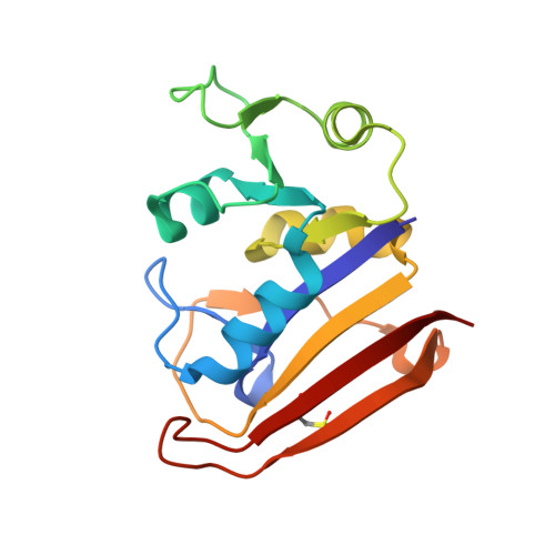
    Single-wavelength anomalous diffraction (SAD) is a routine method for overcoming the phase problem when solving macromolecular structures. This technique requires the accurate measurement of intensities to determine differences between Bijvoet pairs. Although SAD experiments are commonly conducted at cryogenic temperatures to mitigate the effects of radiation damage, such temperatures can alter the conformational ensemble of the protein and may impede the merging of data from multiple crystals due to non-uniform freezing. Here, a strategy is presented to obtain high-quality data from room-temperature, single-crystal experiments. To illustrate the strengths of this approach, native SAD phasing at 6.55 keV was used to solve four structures of three model systems at 295 K. The resulting data sets allow automatic phasing and model building, and reveal alternate conformations that reflect the structure of proteins at room temperature.


  • Organizational Affiliation

    Department of Molecular and Cellular Biology, Harvard University, 52 Oxford Street, Cambridge, Massachusetts, USA.


Macromolecules
Find similar proteins by:  (by identity cutoff)  |  3D Structure
Entity ID: 1
MoleculeChains Sequence LengthOrganismDetailsImage
Dihydrofolate reductase159Escherichia coli K-12Mutation(s): 0 
Gene Names: folAtmrAb0048JW0047
EC: 1.5.1.3
UniProt
Find proteins for P0ABQ4 (Escherichia coli (strain K12))
Explore P0ABQ4 
Go to UniProtKB:  P0ABQ4
Entity Groups  
Sequence Clusters30% Identity50% Identity70% Identity90% Identity95% Identity100% Identity
UniProt GroupP0ABQ4
Sequence Annotations
Expand
  • Reference Sequence
Small Molecules
Modified Residues  1 Unique
IDChains TypeFormula2D DiagramParent
CSD
Query on CSD
A
L-PEPTIDE LINKINGC3 H7 N O4 SCYS
Experimental Data & Validation

Experimental Data

Unit Cell:
Length ( Å )Angle ( ˚ )
a = 34.297α = 90
b = 45.552β = 90
c = 99.035γ = 90
Software Package:
Software NamePurpose
DIALSdata reduction
Aimlessdata scaling
AutoSolphasing
PHENIXmodel building
PHENIXrefinement

Structure Validation

View Full Validation Report


Ligand Structure Quality Assessment 


Entry History & Funding Information

Deposition Data


Funding OrganizationLocationGrant Number
Searle Scholars ProgramUnited StatesSSP-2018-3240
George W. Merck Fund in the New York Community TrustUnited States338034
National Science Foundation (NSF, United States)United StatesDGE1745303

Revision History  (Full details and data files)

  • Version 1.0: 2021-03-17
    Type: Initial release
  • Version 1.1: 2022-08-03
    Changes: Database references
  • Version 1.2: 2022-08-17
    Changes: Database references