6XNN

Crystal Structure of Mouse STING CTD complex with SR-717.


Experimental Data Snapshot

  • Method: X-RAY DIFFRACTION
  • Resolution: 2.49 Å
  • R-Value Free: 0.271 
  • R-Value Work: 0.222 
  • R-Value Observed: 0.225 

wwPDB Validation   3D Report Full Report


Ligand Structure Quality Assessment 


This is version 1.2 of the entry. See complete history


Literature

Antitumor activity of a systemic STING-activating non-nucleotide cGAMP mimetic.

Chin, E.N.Yu, C.Vartabedian, V.F.Jia, Y.Kumar, M.Gamo, A.M.Vernier, W.Ali, S.H.Kissai, M.Lazar, D.C.Nguyen, N.Pereira, L.E.Benish, B.Woods, A.K.Joseph, S.B.Chu, A.Johnson, K.A.Sander, P.N.Martinez-Pena, F.Hampton, E.N.Young, T.S.Wolan, D.W.Chatterjee, A.K.Schultz, P.G.Petrassi, H.M.Teijaro, J.R.Lairson, L.L.

(2020) Science 369: 993-999

  • DOI: https://doi.org/10.1126/science.abb4255
  • Primary Citation of Related Structures:  
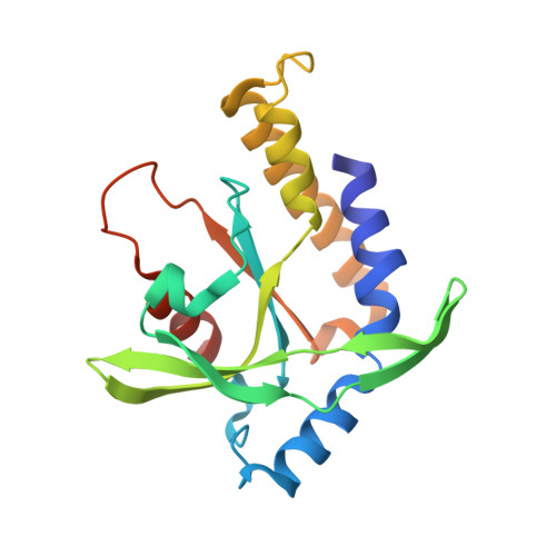
    6XNN, 6XNP

  • PubMed Abstract: 

    Stimulator of interferon genes (STING) links innate immunity to biological processes ranging from antitumor immunity to microbiome homeostasis. Mechanistic understanding of the anticancer potential for STING receptor activation is currently limited by metabolic instability of the natural cyclic dinucleotide (CDN) ligands. From a pathway-targeted cell-based screen, we identified a non-nucleotide, small-molecule STING agonist, termed SR-717, that demonstrates broad interspecies and interallelic specificity. A 1.8-angstrom cocrystal structure revealed that SR-717 functions as a direct cyclic guanosine monophosphate-adenosine monophosphate (cGAMP) mimetic that induces the same "closed" conformation of STING. SR-717 displayed antitumor activity; promoted the activation of CD8 + T, natural killer, and dendritic cells in relevant tissues; and facilitated antigen cross-priming. SR-717 also induced the expression of clinically relevant targets, including programmed cell death 1 ligand 1 (PD-L1), in a STING-dependent manner.


  • Organizational Affiliation

    Department of Chemistry, The Scripps Research Institute, 10550 North Torrey Pines Road, La Jolla, CA 92037, USA.


Macromolecules
Find similar proteins by:  (by identity cutoff)  |  3D Structure
Entity ID: 1
MoleculeChains Sequence LengthOrganismDetailsImage
Stimulator of interferon genes protein
A, B
187Mus musculusMutation(s): 0 
Gene Names: Sting1ErisMitaMpysTmem173
UniProt & NIH Common Fund Data Resources
Find proteins for Q3TBT3 (Mus musculus)
Explore Q3TBT3 
Go to UniProtKB:  Q3TBT3
IMPC:  MGI:1919762
Entity Groups  
Sequence Clusters30% Identity50% Identity70% Identity90% Identity95% Identity100% Identity
UniProt GroupQ3TBT3
Sequence Annotations
Expand
  • Reference Sequence
Small Molecules
Ligands 1 Unique
IDChains Name / Formula / InChI Key2D Diagram3D Interactions
V67 (Subject of Investigation/LOI)
Query on V67

Download Ideal Coordinates CCD File 
C [auth A],
D [auth B]
4,5-difluoro-2-{[6-(1H-imidazol-1-yl)pyridazine-3-carbonyl]amino}benzoic acid
C15 H9 F2 N5 O3
WEBVQIJGIZVRGA-UHFFFAOYSA-N
Experimental Data & Validation

Experimental Data

  • Method: X-RAY DIFFRACTION
  • Resolution: 2.49 Å
  • R-Value Free: 0.271 
  • R-Value Work: 0.222 
  • R-Value Observed: 0.225 
  • Space Group: P 43 21 2
Unit Cell:
Length ( Å )Angle ( ˚ )
a = 65.68α = 90
b = 65.68β = 90
c = 172.577γ = 90
Software Package:
Software NamePurpose
REFMACrefinement
XDSdata reduction
Aimlessdata scaling
PHASERphasing
PDB_EXTRACTdata extraction

Structure Validation

View Full Validation Report


Ligand Structure Quality Assessment 


Entry History 

Deposition Data

Revision History  (Full details and data files)

  • Version 1.0: 2020-08-26
    Type: Initial release
  • Version 1.1: 2020-09-02
    Changes: Database references
  • Version 1.2: 2023-10-18
    Changes: Data collection, Database references, Refinement description