6WFT

BatAAV-10HB - genome-containing particles


Experimental Data Snapshot

  • Method: ELECTRON MICROSCOPY
  • Resolution: 3.03 Å
  • Aggregation State: PARTICLE 
  • Reconstruction Method: SINGLE PARTICLE 

wwPDB Validation   3D Report Full Report


This is version 1.2 of the entry. See complete history


Literature

Structural characterization of a bat Adeno-associated virus capsid.

Mietzsch, M.Li, Y.Kurian, J.Smith, J.K.Chipman, P.McKenna, R.Yang, L.Agbandje-McKenna, M.

(2020) J Struct Biol 211: 107547-107547

  • DOI: https://doi.org/10.1016/j.jsb.2020.107547
  • Primary Citation of Related Structures:  
    6WFT, 6WFU

  • PubMed Abstract: 

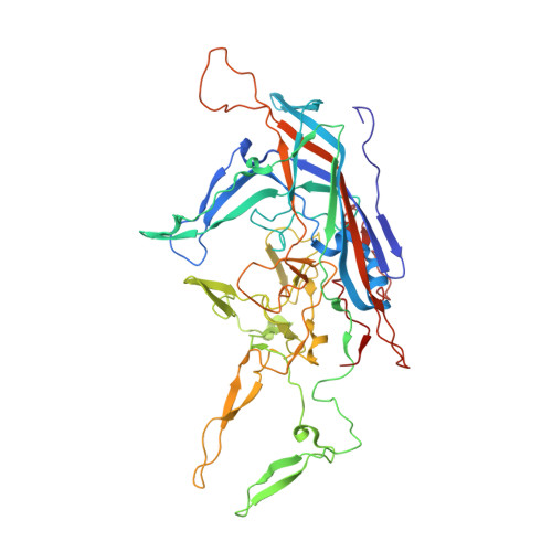
    Adeno-associated viruses (AAVs) are widespread among vertebrates. AAVs isolated from bats display low capsid protein sequence identities (<60%) to AAV2, AAV5, and other primate AAVs. Here we report the first capsid structure of a non-primate AAV which was isolated from bats. The capsid structure of BtAAV-10HB (10HB) was determined by cryo-electron microscopy and three-dimensional image reconstruction to 3.03 Å resolution. Comparison of empty and genome-containing capsids showed that the capsid structures are almost identical except for an ordered nucleotide in a previously described nucleotide-binding pocket, the density in the 5-fold channel, and several amino acids with altered side chain conformations. Compared to other dependoparvoviruses, for example AAV2 and AAV5, 10HB displays unique structural features including insertions and deletions in capsid surface loops. Overall, the 10HB capsid structure superposes with an RMSD of 1.7 Å and 1.8 Å to AAV2 and AAV5, respectively. Currently all approved AAV human gene therapy biologics and vectors in clinical trials are based on primate isolates. However, pre-existing neutralizing antibodies in the human population represents a hurdle to their use. 10HB capsids are capable of packaging AAV2 vector genomes and thus have potential as gene delivery vectors. Significantly, a screen with human sera showed lack of recognition by the 10HB capsid. Thus, the different capsid surface of 10HB vectors likely renders it "invisible" to potential pre-existing neutralizing human anti-AAV antibodies especially because this virus or similar variants do not exist in primate populations.


  • Organizational Affiliation

    Department of Biochemistry and Molecular Biology, Center for Structural Biology, The McKnight Brain Institute, University of Florida, Gainesville, FL, USA.


Macromolecules
Find similar proteins by:  (by identity cutoff)  |  3D Structure
Entity ID: 1
MoleculeChains Sequence LengthOrganismDetailsImage
VP1 capsid513Bat adeno-associated virusMutation(s): 0 
UniProt
Find proteins for A0A2Z4K548 (Bat adeno-associated virus)
Explore A0A2Z4K548 
Go to UniProtKB:  A0A2Z4K548
Entity Groups  
Sequence Clusters30% Identity50% Identity70% Identity90% Identity95% Identity100% Identity
UniProt GroupA0A2Z4K548
Sequence Annotations
Expand
  • Reference Sequence
Small Molecules
Ligands 1 Unique
IDChains Name / Formula / InChI Key2D Diagram3D Interactions
DA
Query on DA

Download Ideal Coordinates CCD File 
AC [auth S]
AD [auth s]
BC [auth T]
BD [auth t]
CC [auth U]
AC [auth S],
AD [auth s],
BC [auth T],
BD [auth t],
CC [auth U],
CD [auth u],
DC [auth V],
DD [auth v],
EC [auth W],
ED [auth w],
FC [auth X],
FD [auth x],
GC [auth Y],
GD [auth y],
HC [auth Z],
HD [auth z],
IB [auth A],
IC [auth a],
ID [auth 1],
JB [auth B],
JC [auth b],
JD [auth 2],
KB [auth C],
KC [auth c],
KD [auth 3],
LB [auth D],
LC [auth d],
LD [auth 4],
MB [auth E],
MC [auth e],
MD [auth 5],
NB [auth F],
NC [auth f],
ND [auth 6],
OB [auth G],
OC [auth g],
OD [auth 7],
PB [auth H],
PC [auth h],
PD [auth 8],
QB [auth I],
QC [auth i],
RB [auth J],
RC [auth j],
SB [auth K],
SC [auth k],
TB [auth L],
TC [auth l],
UB [auth M],
UC [auth m],
VB [auth N],
VC [auth n],
WB [auth O],
WC [auth o],
XB [auth P],
XC [auth p],
YB [auth Q],
YC [auth q],
ZB [auth R],
ZC [auth r]
2'-DEOXYADENOSINE-5'-MONOPHOSPHATE
C10 H14 N5 O6 P
KHWCHTKSEGGWEX-RRKCRQDMSA-N
Experimental Data & Validation

Experimental Data

  • Method: ELECTRON MICROSCOPY
  • Resolution: 3.03 Å
  • Aggregation State: PARTICLE 
  • Reconstruction Method: SINGLE PARTICLE 
EM Software:
TaskSoftware PackageVersion
RECONSTRUCTIONcisTEM

Structure Validation

View Full Validation Report


Entry History & Funding Information

Deposition Data


Funding OrganizationLocationGrant Number
National Institutes of Health/National Institute of General Medical Sciences (NIH/NIGMS)United StatesGM082946

Revision History  (Full details and data files)

  • Version 1.0: 2020-06-24
    Type: Initial release
  • Version 1.1: 2020-07-15
    Changes: Database references
  • Version 1.2: 2024-03-06
    Changes: Data collection, Database references