6VS2

protein D


Experimental Data Snapshot

  • Method: X-RAY DIFFRACTION
  • Resolution: 3.00 Å
  • R-Value Free: 0.299 
  • R-Value Work: 0.280 
  • R-Value Observed: 0.284 

wwPDB Validation   3D Report Full Report


Ligand Structure Quality Assessment 


This is version 1.1 of the entry. See complete history


Literature

Structure and mechanism of a redesigned multidrug transporter from the Major Facilitator Superfamily.

Wu, H.H.Symersky, J.Lu, M.

(2020) Sci Rep 10: 3949-3949

  • DOI: https://doi.org/10.1038/s41598-020-60332-8
  • Primary Citation of Related Structures:  
    6VRZ, 6VS0, 6VS1, 6VS2

  • PubMed Abstract: 

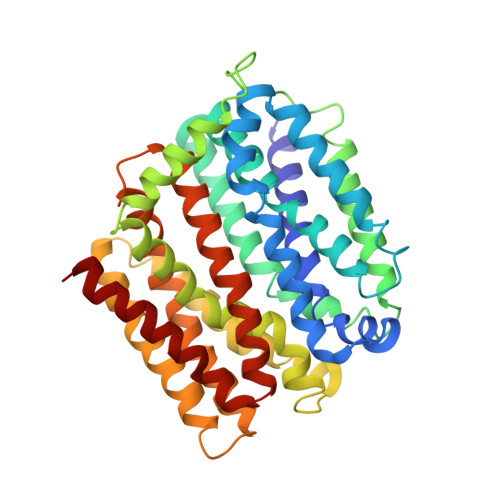
    The rapid increase of multidrug resistance poses urgent threats to human health. Multidrug transporters prompt multidrug resistance by exporting different therapeutics across cell membranes, often by utilizing the H + electrochemical gradient. MdfA from Escherichia coli is a prototypical H + -dependent multidrug transporter belonging to the Major Facilitator Superfamily. Prior studies revealed unusual flexibility in the coupling between multidrug binding and deprotonation in MdfA, but the mechanistic basis for this flexibility was obscure. Here we report the X-ray structures of a MdfA mutant E26T/D34M/A150E, wherein the multidrug-binding and protonation sites were revamped, separately bound to three different substrates at resolutions up to 2.0 Å. To validate the functional relevance of these structures, we conducted mutational and biochemical studies. Our data elucidated intermediate states during antibiotic recognition and suggested structural changes that accompany the substrate-evoked deprotonation of E26T/D34M/A150E. These findings help to explain the mechanistic flexibility in drug/H + coupling observed in MdfA and may inspire therapeutic development to preempt efflux-mediated antimicrobial resistance.


  • Organizational Affiliation

    Center for Proteomics and Molecular Therapeutics, Rosalind Franklin University of Medicine and Science, 3333 Green Bay Road, North Chicago, IL, 60064, USA.


Macromolecules
Find similar proteins by:  (by identity cutoff)  |  3D Structure
Entity ID: 1
MoleculeChains Sequence LengthOrganismDetailsImage
Multidrug transporter MdfA387Escherichia coliMutation(s): 3 
Gene Names: mdfAcmlAcmrb0842JW0826
Membrane Entity: Yes 
UniProt
Find proteins for P0AEY8 (Escherichia coli (strain K12))
Explore P0AEY8 
Go to UniProtKB:  P0AEY8
Entity Groups  
Sequence Clusters30% Identity50% Identity70% Identity90% Identity95% Identity100% Identity
UniProt GroupP0AEY8
Sequence Annotations
Expand
  • Reference Sequence
Small Molecules
Experimental Data & Validation

Experimental Data

  • Method: X-RAY DIFFRACTION
  • Resolution: 3.00 Å
  • R-Value Free: 0.299 
  • R-Value Work: 0.280 
  • R-Value Observed: 0.284 
  • Space Group: C 1 2 1
Unit Cell:
Length ( Å )Angle ( ˚ )
a = 95.014α = 90
b = 66.973β = 111.1
c = 110.433γ = 90
Software Package:
Software NamePurpose
REFMACrefinement
PDB_EXTRACTdata extraction
HKL-2000data reduction
HKL-2000data scaling
SHARPphasing

Structure Validation

View Full Validation Report


Ligand Structure Quality Assessment 


Entry History & Funding Information

Deposition Data


Funding OrganizationLocationGrant Number
National Institutes of Health/National Institute of General Medical Sciences (NIH/NIGMS)United StatesGM094195

Revision History  (Full details and data files)

  • Version 1.0: 2020-06-03
    Type: Initial release
  • Version 1.1: 2024-03-06
    Changes: Advisory, Data collection, Database references