6RLD

STRUCTURE OF THE MECHANOSENSITIVE CHANNEL MSCS EMBEDDED IN THE MEMBRANE BILAYER


Experimental Data Snapshot

  • Method: ELECTRON MICROSCOPY
  • Resolution: 2.90 Å
  • Aggregation State: PARTICLE 
  • Reconstruction Method: SINGLE PARTICLE 

wwPDB Validation   3D Report Full Report


This is version 1.2 of the entry. See complete history


Literature

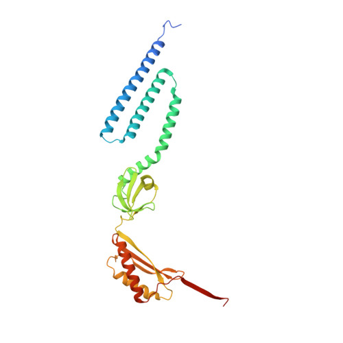
Structure of the Mechanosensitive Channel MscS Embedded in the Membrane Bilayer.

Rasmussen, T.Flegler, V.J.Rasmussen, A.Bottcher, B.

(2019) J Mol Biol 431: 3081-3090

  • DOI: https://doi.org/10.1016/j.jmb.2019.07.006
  • Primary Citation of Related Structures:  
    6RLD

  • PubMed Abstract: 

    Since life has emerged, gradients of osmolytes over the cell membrane cause pressure changes in the cell and require tight regulation to prevent cell rupture. The mechanosensitive channel of small conductance (MscS) releases solutes and water when a hypo-osmotic shock raises the pressure in the cell. It is a member of a large family of MscS-like channels found in bacteria, archaea, fungi and plants and model for mechanosensation. MscS senses the increase of tension in the membrane directly by the force from the lipids, but the molecular mechanism is still elusive. We determined the lipid interactions of MscS by resolving the structure of Escherichia coli MscS embedded in membrane discs to 2.9-Å resolution using cryo-electron microscopy. The membrane is attached only to parts of the sensor paddles of MscS, but phospholipid molecules move through grooves into remote pockets on the cytosolic side. On the periplasmic side, a lipid bound by R88 at the pore entrance is separated from the membrane by TM1 helices. The N-terminus interacts with the periplasmic membrane surface. We demonstrate that the unique membrane domain of MscS promotes deep penetration of lipid molecules and shows multimodal interaction with the membrane to fine-tune tension sensing.


  • Organizational Affiliation

    Biocenter and Rudolf Virchow Center, Universität Würzburg, Haus D15, Josef-Schneider-Str. 2, 97080 Würzburg, Germany. Electronic address: tim.rasmussen@uni-wuerzburg.de.


Macromolecules
Find similar proteins by:  (by identity cutoff)  |  3D Structure
Entity ID: 1
MoleculeChains Sequence LengthOrganismDetailsImage
Small-conductance mechanosensitive channel
A, B, C, D, E
A, B, C, D, E, F, G
286Escherichia coli K-12Mutation(s): 0 
Membrane Entity: Yes 
UniProt
Find proteins for P0C0S1 (Escherichia coli (strain K12))
Explore P0C0S1 
Go to UniProtKB:  P0C0S1
Entity Groups  
Sequence Clusters30% Identity50% Identity70% Identity90% Identity95% Identity100% Identity
UniProt GroupP0C0S1
Sequence Annotations
Expand
  • Reference Sequence
Small Molecules
Ligands 1 Unique
IDChains Name / Formula / InChI Key2D Diagram3D Interactions
PCW (Subject of Investigation/LOI)
Query on PCW

Download Ideal Coordinates CCD File 
AA [auth G]
BA [auth G]
H [auth A]
I [auth A]
J [auth A]
AA [auth G],
BA [auth G],
H [auth A],
I [auth A],
J [auth A],
K [auth B],
L [auth B],
M [auth B],
N [auth C],
O [auth C],
P [auth C],
Q [auth D],
R [auth D],
S [auth D],
T [auth E],
U [auth E],
V [auth E],
W [auth F],
X [auth F],
Y [auth F],
Z [auth G]
1,2-DIOLEOYL-SN-GLYCERO-3-PHOSPHOCHOLINE
C44 H85 N O8 P
SNKAWJBJQDLSFF-NVKMUCNASA-O
Experimental Data & Validation

Experimental Data

  • Method: ELECTRON MICROSCOPY
  • Resolution: 2.90 Å
  • Aggregation State: PARTICLE 
  • Reconstruction Method: SINGLE PARTICLE 
EM Software:
TaskSoftware PackageVersion
MODEL REFINEMENTPHENIX1.13-2998
RECONSTRUCTIONRELION3.0

Structure Validation

View Full Validation Report


Entry History & Funding Information

Deposition Data


Funding OrganizationLocationGrant Number
German Research FoundationGermanyBo1150/15-1
German Research FoundationGermanyINST 93/903-1 FUGG

Revision History  (Full details and data files)

  • Version 1.0: 2019-07-24
    Type: Initial release
  • Version 1.1: 2019-08-21
    Changes: Data collection, Database references
  • Version 1.2: 2019-08-28
    Changes: Data collection, Database references