6R83

CryoEM structure and molecular model of squid hemocyanin (Todarodes pacificus , TpH)


Experimental Data Snapshot

  • Method: ELECTRON MICROSCOPY
  • Resolution: 5.10 Å
  • Aggregation State: PARTICLE 
  • Reconstruction Method: SINGLE PARTICLE 

wwPDB Validation   3D Report Full Report


This is version 1.2 of the entry. See complete history


Literature

Cryo-EM reveals the asymmetric assembly of squid hemocyanin.

Tanaka, Y.Kato, S.Stabrin, M.Raunser, S.Matsui, T.Gatsogiannis, C.

(2019) IUCrJ 6: 426-437

  • DOI: https://doi.org/10.1107/S205225251900321X
  • Primary Citation of Related Structures:  
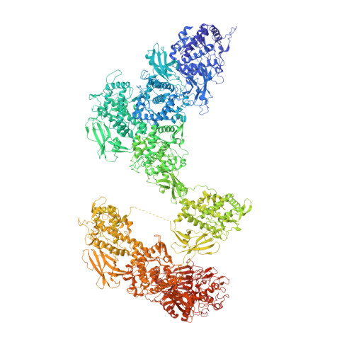
    6R83

  • PubMed Abstract: 

    The oxygen transporter of molluscs, hemocyanin, consists of long pearl-necklace-like subunits of several globular domains. The subunits assemble in a complex manner to form cylindrical decamers. Typically, the first six domains of each subunit assemble together to form the cylinder wall, while the C-terminal domains form a collar that fills or caps the cylinder. During evolution, various molluscs have been able to fine-tune their oxygen binding by deleting or adding C-terminal domains and adjusting their inner-collar architecture. However, squids have duplicated one of the wall domains of their subunits instead. Here, using cryo-EM and an optimized refinement protocol implemented in SPHIRE , this work tackled the symmetry-mismatched structure of squid hemocyanin, revealing the precise effect of this duplication on its quaternary structure and providing a potential model for its structural evolution.


  • Organizational Affiliation

    Graduate School of Life Sciences, Tohoku University, 2-1-1 Katahira, Aoba-ku, Sendai 980-8577, Japan.


Macromolecules
Find similar proteins by:  (by identity cutoff)  |  3D Structure
Entity ID: 1
MoleculeChains Sequence LengthOrganismDetailsImage
Hemocyanin subunit 13,314Todarodes pacificusMutation(s): 0 
UniProt
Find proteins for A0A0P0UX03 (Todarodes pacificus)
Explore A0A0P0UX03 
Go to UniProtKB:  A0A0P0UX03
Entity Groups  
Sequence Clusters30% Identity50% Identity70% Identity90% Identity95% Identity100% Identity
UniProt GroupA0A0P0UX03
Sequence Annotations
Expand
  • Reference Sequence
Experimental Data & Validation

Experimental Data

  • Method: ELECTRON MICROSCOPY
  • Resolution: 5.10 Å
  • Aggregation State: PARTICLE 
  • Reconstruction Method: SINGLE PARTICLE 
EM Software:
TaskSoftware PackageVersion
RECONSTRUCTIONSPHIRE

Structure Validation

View Full Validation Report


Entry History & Funding Information

Deposition Data


Funding OrganizationLocationGrant Number
Max Planck SocietyGermany--
German Research FoundationGermanyFOR1905
Japan Society for the Promotion of ScienceJapan26291008;24000011;15KK0248;16H00748;17K07942

Revision History  (Full details and data files)

  • Version 1.0: 2019-05-08
    Type: Initial release
  • Version 1.1: 2019-05-29
    Changes: Data collection, Database references
  • Version 1.2: 2019-11-06
    Changes: Data collection, Refinement description