6MW8

UDP-galactose:glucoside-Skp1 alpha-D-galactosyltransferase with bound UDP and Manganese


Experimental Data Snapshot

  • Method: X-RAY DIFFRACTION
  • Resolution: 1.76 Å
  • R-Value Free: 0.208 
  • R-Value Work: 0.181 
  • R-Value Observed: 0.182 

wwPDB Validation   3D Report Full Report


Ligand Structure Quality Assessment 


This is version 1.3 of the entry. See complete history


Literature

A terminal alpha 3-galactose modification regulates an E3 ubiquitin ligase subunit in Toxoplasma gondii .

Mandalasi, M.Kim, H.W.Thieker, D.Sheikh, M.O.Gas-Pascual, E.Rahman, K.Zhao, P.Daniel, N.G.van der Wel, H.Ichikawa, H.T.Glushka, J.N.Wells, L.Woods, R.J.Wood, Z.A.West, C.M.

(2020) J Biol Chem 295: 9223-9243

  • DOI: https://doi.org/10.1074/jbc.RA120.013792
  • Primary Citation of Related Structures:  
    6MW5, 6MW8

  • PubMed Abstract: 

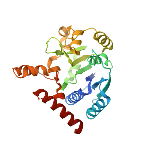
    Skp1, a subunit of E3 Skp1/Cullin-1/F-box protein ubiquitin ligases, is modified by a prolyl hydroxylase that mediates O 2 regulation of the social amoeba Dictyostelium and the parasite Toxoplasma gondii The full effect of hydroxylation requires modification of the hydroxyproline by a pentasaccharide that, in Dictyostelium , influences Skp1 structure to favor assembly of Skp1/F-box protein subcomplexes. In Toxoplasma , the presence of a contrasting penultimate sugar assembled by a different glycosyltransferase enables testing of the conformational control model. To define the final sugar and its linkage, here we identified the glycosyltransferase that completes the glycan and found that it is closely related to glycogenin, an enzyme that may prime glycogen synthesis in yeast and animals. However, the Toxoplasma enzyme catalyzes formation of a Galα1,3Glcα linkage rather than the Glcα1,4Glcα linkage formed by glycogenin. Kinetic and crystallographic experiments showed that the glycosyltransferase Gat1 is specific for Skp1 in Toxoplasma and also in another protist, the crop pathogen Pythium ultimum The fifth sugar is important for glycan function as indicated by the slow-growth phenotype of gat1 Δ parasites. Computational analyses indicated that, despite the sequence difference, the Toxoplasma glycan still assumes an ordered conformation that controls Skp1 structure and revealed the importance of nonpolar packing interactions of the fifth sugar. The substitution of glycosyltransferases in Toxoplasma and Pythium by an unrelated bifunctional enzyme that assembles a distinct but structurally compatible glycan in Dictyostelium is a remarkable case of convergent evolution, which emphasizes the importance of the terminal α-galactose and establishes the phylogenetic breadth of Skp1 glycoregulation.


  • Organizational Affiliation

    Department of Biochemistry and Molecular Biology, University of Georgia, Athens, Georgia, USA; Center for Tropical and Emerging Global Diseases, University of Georgia, Athens, Georgia, USA.


Macromolecules
Find similar proteins by:  (by identity cutoff)  |  3D Structure
Entity ID: 1
MoleculeChains Sequence LengthOrganismDetailsImage
UDP-galactose:glucoside-Skp1 alpha-D-galactosyltransferase266Globisporangium ultimumMutation(s): 0 
EC: 2.4.1.87
UniProt
Find proteins for K3WC47 (Globisporangium ultimum (strain ATCC 200006 / CBS 805.95 / DAOM BR144))
Explore K3WC47 
Go to UniProtKB:  K3WC47
Entity Groups  
Sequence Clusters30% Identity50% Identity70% Identity90% Identity95% Identity100% Identity
UniProt GroupK3WC47
Sequence Annotations
Expand
  • Reference Sequence
Small Molecules
Ligands 4 Unique
IDChains Name / Formula / InChI Key2D Diagram3D Interactions
UDP (Subject of Investigation/LOI)
Query on UDP

Download Ideal Coordinates CCD File 
C [auth A]URIDINE-5'-DIPHOSPHATE
C9 H14 N2 O12 P2
XCCTYIAWTASOJW-XVFCMESISA-N
EDO
Query on EDO

Download Ideal Coordinates CCD File 
G [auth A]1,2-ETHANEDIOL
C2 H6 O2
LYCAIKOWRPUZTN-UHFFFAOYSA-N
ACT
Query on ACT

Download Ideal Coordinates CCD File 
D [auth A],
E [auth A],
F [auth A]
ACETATE ION
C2 H3 O2
QTBSBXVTEAMEQO-UHFFFAOYSA-M
MN (Subject of Investigation/LOI)
Query on MN

Download Ideal Coordinates CCD File 
B [auth A]MANGANESE (II) ION
Mn
WAEMQWOKJMHJLA-UHFFFAOYSA-N
Experimental Data & Validation

Experimental Data

  • Method: X-RAY DIFFRACTION
  • Resolution: 1.76 Å
  • R-Value Free: 0.208 
  • R-Value Work: 0.181 
  • R-Value Observed: 0.182 
  • Space Group: P 42 21 2
Unit Cell:
Length ( Å )Angle ( ˚ )
a = 84.055α = 90
b = 84.055β = 90
c = 76.076γ = 90
Software Package:
Software NamePurpose
XDSdata scaling
XDSdata reduction
PHENIXphasing
PHENIXrefinement
Cootmodel building

Structure Validation

View Full Validation Report


Ligand Structure Quality Assessment 


Entry History & Funding Information

Deposition Data


Funding OrganizationLocationGrant Number
National Institutes of Health/National Institute of General Medical Sciences (NIH/NIGMS)United StatesR01-GM084383

Revision History  (Full details and data files)

  • Version 1.0: 2019-10-09
    Type: Initial release
  • Version 1.1: 2020-01-01
    Changes: Author supporting evidence
  • Version 1.2: 2020-10-21
    Changes: Database references
  • Version 1.3: 2024-03-13
    Changes: Data collection, Database references