6KZA

Crystal structure of the complex of the interaction domains of E. coli DnaB helicase and DnaC helicase loader


Experimental Data Snapshot

  • Method: X-RAY DIFFRACTION
  • Resolution: 3.10 Å
  • R-Value Free: 0.267 
  • R-Value Work: 0.232 
  • R-Value Observed: 0.234 

wwPDB Validation   3D Report Full Report


This is version 1.2 of the entry. See complete history


Literature

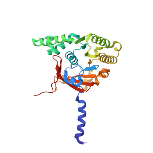
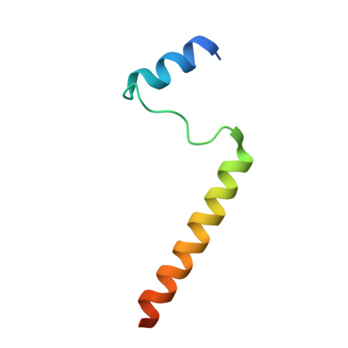
Crystal structure of the complex of the interaction domains of Escherichia coli DnaB helicase and DnaC helicase loader: structural basis implying a distortion-accumulation mechanism for the DnaB ring opening caused by DnaC binding.

Nagata, K.Okada, A.Ohtsuka, J.Ohkuri, T.Akama, Y.Sakiyama, Y.Miyazaki, E.Horita, S.Katayama, T.Ueda, T.Tanokura, M.

(2020) J Biochem 167: 1-14

  • DOI: https://doi.org/10.1093/jb/mvz087
  • Primary Citation of Related Structures:  
    6KZA

  • PubMed Abstract: 

    Loading the bacterial replicative helicase DnaB onto DNA requires a specific loader protein, DnaC/DnaI, which creates the loading-competent state by opening the DnaB hexameric ring. To understand the molecular mechanism by which DnaC/DnaI opens the DnaB ring, we solved 3.1-Å co-crystal structure of the interaction domains of Escherichia coli DnaB-DnaC. The structure reveals that one N-terminal domain (NTD) of DnaC interacts with both the linker helix of a DnaB molecule and the C-terminal domain (CTD) of the adjacent DnaB molecule by forming a three α-helix bundle, which fixes the relative orientation of the two adjacent DnaB CTDs. The importance of the intermolecular interface in the crystal structure was supported by the mutational data of DnaB and DnaC. Based on the crystal structure and other available information on DnaB-DnaC structures, we constructed a molecular model of the hexameric DnaB CTDs bound by six DnaC NTDs. This model suggested that the binding of a DnaC would cause a distortion in the hexameric ring of DnaB. This distortion of the DnaB ring might accumulate by the binding of up to six DnaC molecules, resulting in the DnaB ring to open.


  • Organizational Affiliation

    Department of Applied Biological Chemistry, Graduate School of Agricultural and Life Sciences, The University of Tokyo, Bunkyo-ku, Tokyo, Japan.


Macromolecules
Find similar proteins by:  (by identity cutoff)  |  3D Structure
Entity ID: 1
MoleculeChains Sequence LengthOrganismDetailsImage
Replicative DNA helicase
A, B
292Escherichia coliMutation(s): 0 
Gene Names: dnaBEVY21_06600
EC: 3.6.4.12
UniProt
Find proteins for P0ACB0 (Escherichia coli (strain K12))
Explore P0ACB0 
Go to UniProtKB:  P0ACB0
Entity Groups  
Sequence Clusters30% Identity50% Identity70% Identity90% Identity95% Identity100% Identity
UniProt GroupP0ACB0
Sequence Annotations
Expand
  • Reference Sequence
Find similar proteins by:  (by identity cutoff)  |  3D Structure
Entity ID: 2
MoleculeChains Sequence LengthOrganismDetailsImage
DNA replication protein DnaC
C, D
56Escherichia coliMutation(s): 0 
Gene Names: dnaCEYX85_09815EYY00_08890EYY22_09440
UniProt
Find proteins for P0AEF0 (Escherichia coli (strain K12))
Explore P0AEF0 
Go to UniProtKB:  P0AEF0
Entity Groups  
Sequence Clusters30% Identity50% Identity70% Identity90% Identity95% Identity100% Identity
UniProt GroupP0AEF0
Sequence Annotations
Expand
  • Reference Sequence
Experimental Data & Validation

Experimental Data

  • Method: X-RAY DIFFRACTION
  • Resolution: 3.10 Å
  • R-Value Free: 0.267 
  • R-Value Work: 0.232 
  • R-Value Observed: 0.234 
  • Space Group: P 42 21 2
Unit Cell:
Length ( Å )Angle ( ˚ )
a = 121.56α = 90
b = 121.56β = 90
c = 105.55γ = 90
Software Package:
Software NamePurpose
XDSdata reduction
Aimlessdata scaling
MOLREPphasing
REFMACrefinement
PDB_EXTRACTdata extraction

Structure Validation

View Full Validation Report


Entry History & Funding Information

Deposition Data


Funding OrganizationLocationGrant Number
Japan Society for the Promotion of ScienceJapan26291004
Japan Society for the Promotion of ScienceJapan22-6606
Ministry of Education, Culture, Sports, Science and Technology (Japan)JapanTPRP
Japan Agency for Medical Research and Development (AMED)JapanPDIS

Revision History  (Full details and data files)

  • Version 1.0: 2019-11-20
    Type: Initial release
  • Version 1.1: 2020-01-22
    Changes: Database references
  • Version 1.2: 2024-03-27
    Changes: Data collection, Database references