6KCN

Crystal structure of plasmodium lysyl-tRNA synthetase in complex with a cladosporin derivative 4


Experimental Data Snapshot

  • Method: X-RAY DIFFRACTION
  • Resolution: 2.20 Å
  • R-Value Free: 0.233 
  • R-Value Work: 0.188 
  • R-Value Observed: 0.191 

wwPDB Validation   3D Report Full Report


Ligand Structure Quality Assessment 


This is version 1.2 of the entry. See complete history


Literature

Atomic Resolution Analyses of Isocoumarin Derivatives for Inhibition of Lysyl-tRNA Synthetase.

Zhou, J.Zheng, L.Hei, Z.Li, W.Wang, J.Yu, B.Fang, P.

(2020) ACS Chem Biol 15: 1016-1025

  • DOI: https://doi.org/10.1021/acschembio.0c00032
  • Primary Citation of Related Structures:  
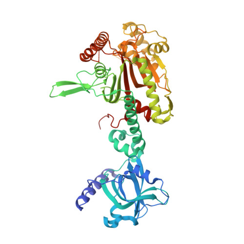
    6KA6, 6KAB, 6KBF, 6KCN, 6KCT

  • PubMed Abstract: 

    Aminoacyl-tRNA synthetases, the essential enzyme family for protein translation, are attractive targets for developing antibacterial, antifungal, and antiparasitic agents and for treating other human diseases. The antimalarial natural product cladosporin was discovered recently as a novel lysyl-tRNA synthetase (LysRS) specific inhibitor. Here, we report a thorough analysis of cladosporin derivatives using chemical synthesis, biophysical, and biochemical experiments. A series of isocoumarin derivatives with only one nonhydrogen atom/bond change per compound was synthesized. These changes include replacements of methyltetrahydropyran moiety by methylcyclohexane or cyclohexane, lactone by lactam, hydroxyl groups by methoxyl groups, and dismission of the chiral center at C3 with a Δ 3,4 double bond. We evaluated these compounds by thermal shift assays and enzymatic experiments and further studied their molecular recognition by the Plasmodium falciparum LysRS through total five high-resolution crystal structures. Our results showed that the methyltetrahydropyran moiety of cladosporin could be replaced by a more stable methylcyclohexane without reducing binding ability. Removing the methyl group from the methylcyclohexane moiety slightly decreased the interaction with LysRS. Besides, the replacement with a lactam group or a conjugated Δ 3,4 double bond within the scaffold could be two more options to optimize the compound. Lastly, the two phenolic hydroxyl groups were critical for the compounds to bind LysRS. The detailed analyses at atomic resolution in this study provide a foundation for the further development of new antibiotics from cladosporin derivatives.


  • Organizational Affiliation

    State Key Laboratory of Bioorganic and Natural Products Chemistry, Center for Excellence in Molecular Synthesis, Shanghai Institute of Organic Chemistry, University of Chinese Academy of Sciences, Chinese Academy of Sciences, 345 Lingling Road, Shanghai 200032, China.


Macromolecules
Find similar proteins by:  (by identity cutoff)  |  3D Structure
Entity ID: 1
MoleculeChains Sequence LengthOrganismDetailsImage
Lysine--tRNA ligase
A, B, C, D
516Plasmodium falciparum NF54Mutation(s): 0 
Gene Names: PFNF54_04763
EC: 6.1.1.6
UniProt
Find proteins for W7JP72 (Plasmodium falciparum (isolate NF54))
Explore W7JP72 
Go to UniProtKB:  W7JP72
Entity Groups  
Sequence Clusters30% Identity50% Identity70% Identity90% Identity95% Identity100% Identity
UniProt GroupW7JP72
Sequence Annotations
Expand
  • Reference Sequence
Small Molecules
Ligands 3 Unique
IDChains Name / Formula / InChI Key2D Diagram3D Interactions
D5F (Subject of Investigation/LOI)
Query on D5F

Download Ideal Coordinates CCD File 
E [auth A],
H [auth B],
K [auth C],
O [auth D]
3-[[(1~{S},3~{S})-3-methylcyclohexyl]methyl]-6,8-bis(oxidanyl)isochromen-1-one
C17 H20 O4
JSPLICPLSKXJBM-QWRGUYRKSA-N
LYS
Query on LYS

Download Ideal Coordinates CCD File 
G [auth A],
J [auth B],
N [auth C],
P [auth D]
LYSINE
C6 H15 N2 O2
KDXKERNSBIXSRK-YFKPBYRVSA-O
GOL
Query on GOL

Download Ideal Coordinates CCD File 
F [auth A],
I [auth B],
L [auth C],
M [auth C]
GLYCEROL
C3 H8 O3
PEDCQBHIVMGVHV-UHFFFAOYSA-N
Binding Affinity Annotations 
IDSourceBinding Affinity
D5F Binding MOAD:  6KCN IC50: 276 (nM) from 1 assay(s)
Experimental Data & Validation

Experimental Data

  • Method: X-RAY DIFFRACTION
  • Resolution: 2.20 Å
  • R-Value Free: 0.233 
  • R-Value Work: 0.188 
  • R-Value Observed: 0.191 
  • Space Group: P 1
Unit Cell:
Length ( Å )Angle ( ˚ )
a = 71.174α = 76.7
b = 89.92β = 72.95
c = 100.044γ = 80.11
Software Package:
Software NamePurpose
PHENIXrefinement
HKL-2000data scaling
PDB_EXTRACTdata extraction
HKL-3000data reduction
MOLREPphasing

Structure Validation

View Full Validation Report


Ligand Structure Quality Assessment 


Entry History & Funding Information

Deposition Data


Funding OrganizationLocationGrant Number
National Science Foundation (China)China21778064

Revision History  (Full details and data files)

  • Version 1.0: 2020-04-08
    Type: Initial release
  • Version 1.1: 2020-05-06
    Changes: Database references
  • Version 1.2: 2023-11-22
    Changes: Data collection, Database references, Refinement description