6GTY

Crystal structure of the FimH lectin domain from E.coli K12 in complex with the dimannoside Man(alpha1-6)Man


Experimental Data Snapshot

  • Method: X-RAY DIFFRACTION
  • Resolution: 1.90 Å
  • R-Value Free: 0.216 
  • R-Value Work: 0.182 
  • R-Value Observed: 0.184 

wwPDB Validation   3D Report Full Report


This is version 2.1 of the entry. See complete history


Literature

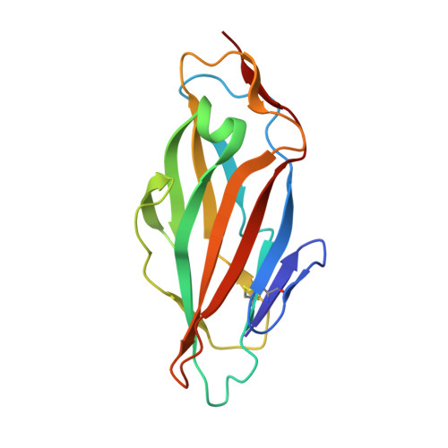
Binding of the Bacterial Adhesin FimH to Its Natural, Multivalent High-Mannose Type Glycan Targets.

Sauer, M.M.Jakob, R.P.Luber, T.Canonica, F.Navarra, G.Ernst, B.Unverzagt, C.Maier, T.Glockshuber, R.

(2019) J Am Chem Soc 141: 936-944

  • DOI: https://doi.org/10.1021/jacs.8b10736
  • Primary Citation of Related Structures:  
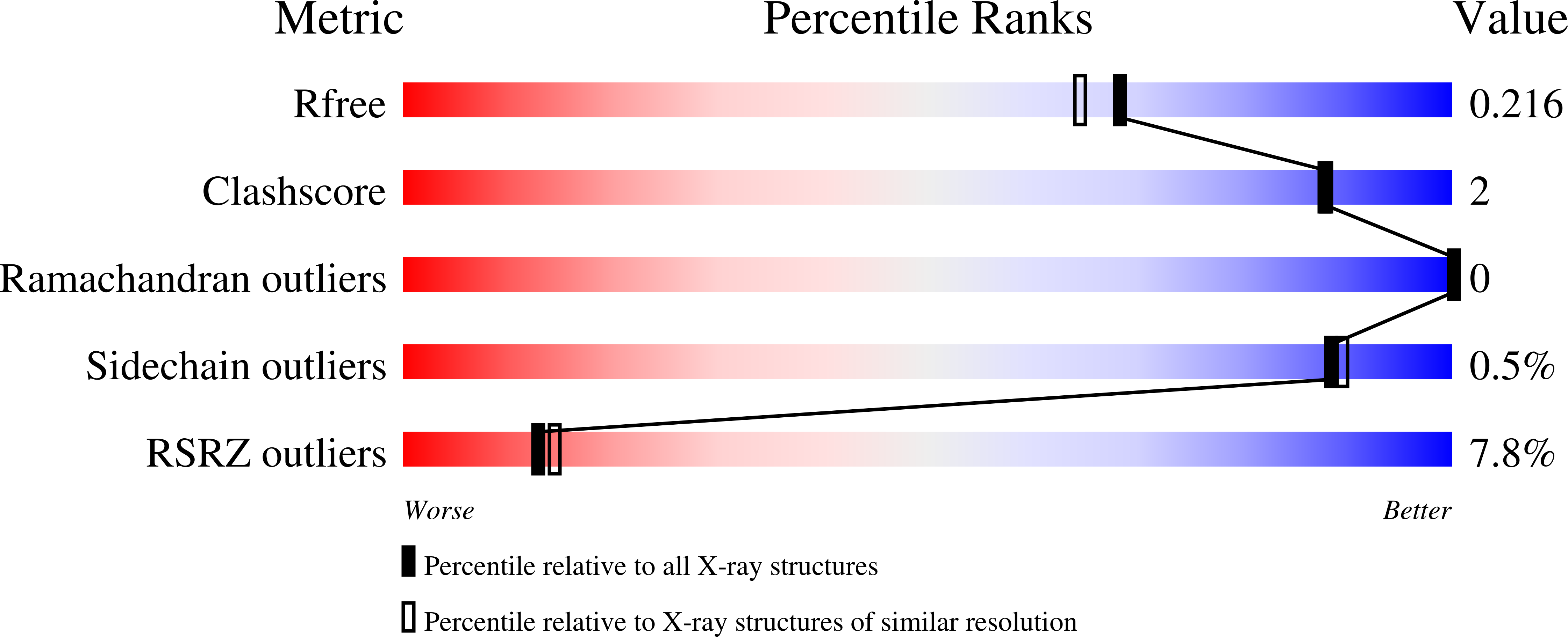
    6GTV, 6GTW, 6GTX, 6GTY, 6GTZ, 6GU0

  • PubMed Abstract: 

    Multivalent carbohydrate-lectin interactions at host-pathogen interfaces play a crucial role in the establishment of infections. Although competitive antagonists that prevent pathogen adhesion are promising antimicrobial drugs, the molecular mechanisms underlying these complex adhesion processes are still poorly understood. Here, we characterize the interactions between the fimbrial adhesin FimH from uropathogenic Escherichia coli strains and its natural high-mannose type N-glycan binding epitopes on uroepithelial glycoproteins. Crystal structures and a detailed kinetic characterization of ligand-binding and dissociation revealed that the binding pocket of FimH evolved such that it recognizes the terminal α(1-2)-, α(1-3)-, and α(1-6)-linked mannosides of natural high-mannose type N-glycans with similar affinity. We demonstrate that the 2000-fold higher affinity of the domain-separated state of FimH compared to its domain-associated state is ligand-independent and consistent with a thermodynamic cycle in which ligand-binding shifts the association equilibrium between the FimH lectin and the FimH pilin domain. Moreover, we show that a single N-glycan can bind up to three molecules of FimH, albeit with negative cooperativity, so that a molar excess of accessible N-glycans over FimH on the cell surface favors monovalent FimH binding. Our data provide pivotal insights into the adhesion properties of uropathogenic Escherichia coli strains to their target receptors and a solid basis for the development of effective FimH antagonists.


  • Organizational Affiliation

    Institute of Molecular Biology & Biophysics , ETH Zurich , Otto-Stern-Weg 5 , CH-8093 Zurich , Switzerland.


Macromolecules
Find similar proteins by:  (by identity cutoff)  |  3D Structure
Entity ID: 1
MoleculeChains Sequence LengthOrganismDetailsImage
Type 1 fimbrin D-mannose specific adhesin
A, B, C, D, E
158Escherichia coli K-12Mutation(s): 0 
Gene Names: fimHb4320JW4283
UniProt
Find proteins for P08191 (Escherichia coli (strain K12))
Explore P08191 
Go to UniProtKB:  P08191
Entity Groups  
Sequence Clusters30% Identity50% Identity70% Identity90% Identity95% Identity100% Identity
UniProt GroupP08191
Sequence Annotations
Expand
  • Reference Sequence
Oligosaccharides

Help

Entity ID: 2
MoleculeChains Length2D Diagram Glycosylation3D Interactions
alpha-D-mannopyranose-(1-6)-methyl alpha-D-mannopyranoside
F, G, H, I, J
2N/A
Glycosylation Resources
GlyTouCan:  G76634EM
GlyCosmos:  G76634EM
Experimental Data & Validation

Experimental Data

  • Method: X-RAY DIFFRACTION
  • Resolution: 1.90 Å
  • R-Value Free: 0.216 
  • R-Value Work: 0.182 
  • R-Value Observed: 0.184 
  • Space Group: P 43
Unit Cell:
Length ( Å )Angle ( ˚ )
a = 90.61α = 90
b = 90.61β = 90
c = 91.53γ = 90
Software Package:
Software NamePurpose
PHENIXrefinement
XDSdata reduction
XSCALEdata scaling
PHASERphasing

Structure Validation

View Full Validation Report


Entry History 

Deposition Data

Revision History  (Full details and data files)

  • Version 1.0: 2019-01-16
    Type: Initial release
  • Version 1.1: 2019-05-01
    Changes: Data collection, Database references
  • Version 2.0: 2020-07-29
    Type: Remediation
    Reason: Carbohydrate remediation
    Changes: Atomic model, Data collection, Derived calculations, Structure summary
  • Version 2.1: 2024-01-17
    Changes: Data collection, Database references, Derived calculations, Refinement description, Structure summary