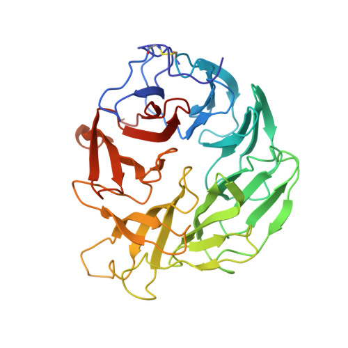
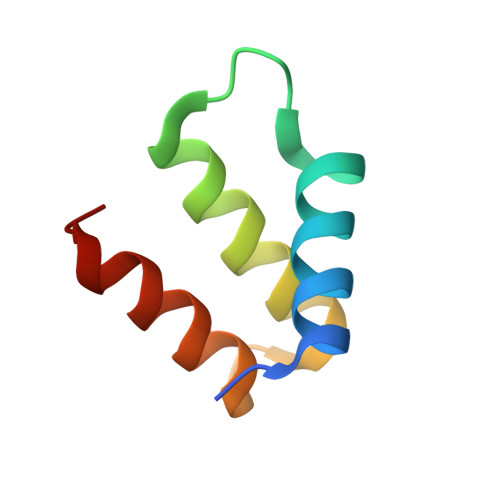
Destructive twisting of neutral metalloproteases: the catalysis mechanism of the Dispase autolysis-inducing protein from Streptomyces mobaraensis DSM 40487.
Fiebig, D., Storka, J., Roeder, M., Meyners, C., Schmelz, S., Blankenfeldt, W., Scrima, A., Kolmar, H., Fuchsbauer, H.L.(2018) FEBS J 285: 4246-4264
- PubMed: 30171661
- DOI: https://doi.org/10.1111/febs.14647
- Primary Citation of Related Structures:
6FHP - PubMed Abstract:
The Dispase autolysis-inducing protein (DAIP) is produced by Streptomyces mobaraensis to disarm neutral metalloproteases by decomposition. The absence of a catalytic protease domain led to the assumption that the seven-bladed β-propeller protein DAIP causes structural modifications, thereby triggering autolysis. Determination of protein complexes consisting of DAIP and thermolysin or DAIP and a nonfunctional E138A bacillolysin variant supported this postulation. Protein twisting was indicated by DAIP-mediated inhibition of thermolysin while bacillolysin underwent immediate autolysis under the same conditions. Interestingly, an increase in SYPRO orange fluorescence allowed tracking of the fast degradation process. Similarly rapid autolysis of thermolysin mediated by DAIP was only observed upon the addition of amphiphilic compounds, which probably amplify the induced structural changes. DAIP further caused degradation of FITC-labeled E138A bacillolysin by trypsin, as monitored by a linear decrease in fluorescence polarization. The kinetic model, calculated from the obtained data, suggested a three-step mechanism defined by (a) fast DAIP-metalloprotease complex formation, (b) slower DAIP-mediated protein twisting, and (c) fragmentation. These results were substantiated by crystallized DAIP attached to a C-terminal helix fragment of thermolysin. Structural superposition of the complex with thermolysin is indicative of a conformational change upon binding to DAIP. Importantly, the majority of metalloproteases, also including homologs from various pathogens, are highly conserved at the autolysis-prone peptide bonds, suggesting their susceptibility to DAIP-mediated decomposition, which may offer opportunities for pharmaceutical applications. DATABASES: The atomic coordinates and structure factors (PDB ID: 6FHP) have been deposited in the Protein Data Bank (http://www.pdb.org/). ENZYMES: Aureolysin, EC 3.4.24.29; bacillolysin (Dispase, Gentlyase), EC 3.4.24.28; lasB (elastase), EC 3.4.24.4; subtilisin, EC 3.4.21.62; thermolysin, EC 3.4.24.27; transglutaminase, EC 2.3.2.13; trypsin, EC 3.4.21.4; vibriolysin (hemagglutinin(HA)/protease), EC 3.4.24.25.
Organizational Affiliation:
Department of Chemical Engineering and Biotechnology, University of Applied Sciences of Darmstadt, Germany.