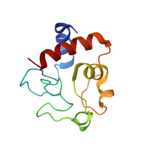
6FF5

X-ray structure of bovine heart cytochrome c at high ionic strength


Experimental Data Snapshot

  • Method: X-RAY DIFFRACTION
  • Resolution: 1.74 Å
  • R-Value Free: 0.203 
  • R-Value Work: 0.161 
  • R-Value Observed: 0.163 

wwPDB Validation   3D Report Full Report


Ligand Structure Quality Assessment 


This is version 1.2 of the entry. See complete history


Literature

X-ray structure of bovine heart cytochrome c at high ionic strength.

Merlino, A.

(2018) Biometals 31: 277-284

  • DOI: https://doi.org/10.1007/s10534-018-0090-x
  • Primary Citation of Related Structures:  
    6FF5

  • PubMed Abstract: 

    Bovine heart cytochrome c (bCyt c) is an extensively studied hemoprotein of only 104 residues. Due to the existence of isoforms generated by non-enzymatic deaminidation, crystallization of bCyt c is difficult and involves extensive purification and the use of microseeding or the presence of an electric field. Taking advantage of the capacity of cytochrome c (cyt c) to bind anions on its protein surface, the commercially available bCyt c was crystallized without extra purifications, using ammonium sulfate as precipitant and nitrate ions as additives. The structure of the ferric bCyt c in a new crystal form is described and compared with that previously solved at low ionic strength and with those of human and horse cyt c. The overall structure of bCyt c is conserved, while the side chains of several residues that play a role in the interactions of cyt c with its partners have different rotamers in the two structures. The effect of the presence of nitrate ions on the structure of the protein is then evaluated and compared with that observed in the case of ferrous and ferric horse heart cyt c.


  • Organizational Affiliation

    Department of Chemical Sciences, University of Naples Federico II, Complesso Universitario di Monte Sant'Angelo, Via Cintia, 80126, Naples, Italy. antonello.merlino@unina.it.


Macromolecules
Find similar proteins by:  (by identity cutoff)  |  3D Structure
Entity ID: 1
MoleculeChains Sequence LengthOrganismDetailsImage
Cytochrome c104Bos taurusMutation(s): 0 
UniProt
Find proteins for P62894 (Bos taurus)
Explore P62894 
Go to UniProtKB:  P62894
Entity Groups  
Sequence Clusters30% Identity50% Identity70% Identity90% Identity95% Identity100% Identity
UniProt GroupP62894
Sequence Annotations
Expand
  • Reference Sequence
Experimental Data & Validation

Experimental Data

  • Method: X-RAY DIFFRACTION
  • Resolution: 1.74 Å
  • R-Value Free: 0.203 
  • R-Value Work: 0.161 
  • R-Value Observed: 0.163 
  • Space Group: H 3 2
Unit Cell:
Length ( Å )Angle ( ˚ )
a = 83.35α = 90
b = 83.35β = 90
c = 92.49γ = 120
Software Package:
Software NamePurpose
REFMACrefinement
XDSdata reduction
SCALAdata scaling
PHASERphasing

Structure Validation

View Full Validation Report


Ligand Structure Quality Assessment 


Entry History 

Deposition Data

  • Released Date: 2018-03-21 
  • Deposition Author(s): Merlino, A.

Revision History  (Full details and data files)

  • Version 1.0: 2018-03-21
    Type: Initial release
  • Version 1.1: 2018-04-04
    Changes: Data collection, Database references
  • Version 1.2: 2024-01-17
    Changes: Data collection, Database references, Refinement description