6ES0

Crystal structure of the kinase domain of human RIPK2 in complex with the activation loop targeting inhibitor CS-R35


Experimental Data Snapshot

  • Method: X-RAY DIFFRACTION
  • Resolution: 2.38 Å
  • R-Value Free: 0.258 
  • R-Value Work: 0.213 
  • R-Value Observed: 0.216 

wwPDB Validation   3D Report Full Report


Ligand Structure Quality Assessment 


This is version 1.3 of the entry. See complete history


Literature

Activation loop targeting strategy for design of receptor-interacting protein kinase 2 (RIPK2) inhibitors.

Suebsuwong, C.Pinkas, D.M.Ray, S.S.Bufton, J.C.Dai, B.Bullock, A.N.Degterev, A.Cuny, G.D.

(2018) Bioorg Med Chem Lett 28: 577-583

  • DOI: https://doi.org/10.1016/j.bmcl.2018.01.044
  • Primary Citation of Related Structures:  
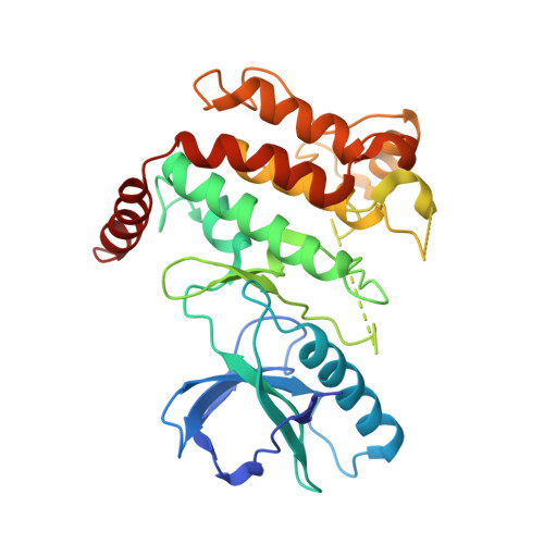
    6ES0

  • PubMed Abstract: 

    Development of selective kinase inhibitors remains a challenge due to considerable amino acid sequence similarity among family members particularly in the ATP binding site. Targeting the activation loop might offer improved inhibitor selectivity since this region of kinases is less conserved. However, the strategy presents difficulties due to activation loop flexibility. Herein, we report the design of receptor-interacting protein kinase 2 (RIPK2) inhibitors based on pan-kinase inhibitor regorafenib that aim to engage basic activation loop residues Lys169 or Arg171. We report development of CSR35 that displayed >10-fold selective inhibition of RIPK2 versus VEGFR2, the target of regorafenib. A co-crystal structure of CSR35 with RIPK2 revealed a resolved activation loop with an ionic interaction between the carboxylic acid installed in the inhibitor and the side-chain of Lys169. Our data provides principle feasibility of developing activation loop targeting type II inhibitors as a complementary strategy for achieving improved selectivity.


  • Organizational Affiliation

    Department of Chemistry, University of Houston, Science and Research Building 2, Houston, TX 77204, USA.


Macromolecules
Find similar proteins by:  (by identity cutoff)  |  3D Structure
Entity ID: 1
MoleculeChains Sequence LengthOrganismDetailsImage
Receptor-interacting serine/threonine-protein kinase 2
A, B
317Homo sapiensMutation(s): 0 
Gene Names: RIPK2CARDIAKRICKRIP2UNQ277/PRO314/PRO34092
EC: 2.7.11.1 (PDB Primary Data), 2.7.10.2 (PDB Primary Data)
UniProt & NIH Common Fund Data Resources
Find proteins for O43353 (Homo sapiens)
Explore O43353 
Go to UniProtKB:  O43353
PHAROS:  O43353
GTEx:  ENSG00000104312 
Entity Groups  
Sequence Clusters30% Identity50% Identity70% Identity90% Identity95% Identity100% Identity
UniProt GroupO43353
Sequence Annotations
Expand
  • Reference Sequence
Small Molecules
Ligands 1 Unique
IDChains Name / Formula / InChI Key2D Diagram3D Interactions
BW8 (Subject of Investigation/LOI)
Query on BW8

Download Ideal Coordinates CCD File 
C [auth A],
D [auth B]
2-[2-fluoranyl-4-[[2-fluoranyl-4-[2-(methylcarbamoyl)pyridin-4-yl]oxy-phenyl]carbamoylamino]phenyl]sulfanylethanoic acid
C22 H18 F2 N4 O5 S
LUCVAGIVDZCQRA-UHFFFAOYSA-N
Binding Affinity Annotations 
IDSourceBinding Affinity
BW8 BindingDB:  6ES0 IC50: min: 2260, max: 4870 (nM) from 2 assay(s)
Binding MOAD:  6ES0 IC50: 2260 (nM) from 1 assay(s)
Experimental Data & Validation

Experimental Data

  • Method: X-RAY DIFFRACTION
  • Resolution: 2.38 Å
  • R-Value Free: 0.258 
  • R-Value Work: 0.213 
  • R-Value Observed: 0.216 
  • Space Group: P 21 21 21
Unit Cell:
Length ( Å )Angle ( ˚ )
a = 61.98α = 90
b = 83.74β = 90
c = 139.1γ = 90
Software Package:
Software NamePurpose
PHENIXrefinement
XDSdata reduction
X-Areadata scaling
PHASERphasing

Structure Validation

View Full Validation Report


Ligand Structure Quality Assessment 


Entry History 

Revision History  (Full details and data files)

  • Version 1.0: 2018-02-21
    Type: Initial release
  • Version 1.1: 2018-02-28
    Changes: Database references
  • Version 1.2: 2019-10-16
    Changes: Data collection
  • Version 1.3: 2024-01-17
    Changes: Data collection, Database references, Refinement description