6EHL

Model of the Ebola virus nucleoprotein in recombinant nucleocapsid-like assemblies


Experimental Data Snapshot

  • Method: ELECTRON MICROSCOPY
  • Resolution: 6.60 Å
  • Aggregation State: HELICAL ARRAY 
  • Reconstruction Method: SUBTOMOGRAM AVERAGING 

wwPDB Validation   3D Report Full Report


This is version 1.4 of the entry. See complete history


Literature

Structure and assembly of the Ebola virus nucleocapsid.

Wan, W.Kolesnikova, L.Clarke, M.Koehler, A.Noda, T.Becker, S.Briggs, J.A.G.

(2017) Nature 551: 394-397

  • DOI: https://doi.org/10.1038/nature24490
  • Primary Citation of Related Structures:  
    6EHL, 6EHM

  • PubMed Abstract: 

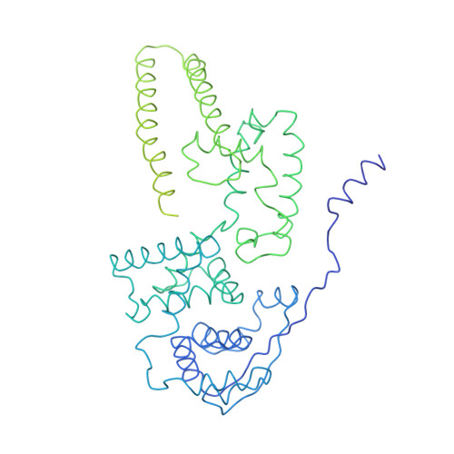
    Ebola and Marburg viruses are filoviruses: filamentous, enveloped viruses that cause haemorrhagic fever. Filoviruses are within the order Mononegavirales, which also includes rabies virus, measles virus, and respiratory syncytial virus. Mononegaviruses have non-segmented, single-stranded negative-sense RNA genomes that are encapsidated by nucleoprotein and other viral proteins to form a helical nucleocapsid. The nucleocapsid acts as a scaffold for virus assembly and as a template for genome transcription and replication. Insights into nucleoprotein-nucleoprotein interactions have been derived from structural studies of oligomerized, RNA-encapsidating nucleoprotein, and cryo-electron microscopy of nucleocapsid or nucleocapsid-like structures. There have been no high-resolution reconstructions of complete mononegavirus nucleocapsids. Here we apply cryo-electron tomography and subtomogram averaging to determine the structure of Ebola virus nucleocapsid within intact viruses and recombinant nucleocapsid-like assemblies. These structures reveal the identity and arrangement of the nucleocapsid components, and suggest that the formation of an extended α-helix from the disordered carboxy-terminal region of nucleoprotein-core links nucleoprotein oligomerization, nucleocapsid condensation, RNA encapsidation, and accessory protein recruitment.


  • Organizational Affiliation

    Structural and Computational Biology Unit, European Molecular Biology Laboratory, Meyerhofstraße 1, 69117 Heidelberg, Germany.


Macromolecules
Find similar proteins by:  (by identity cutoff)  |  3D Structure
Entity ID: 1
MoleculeChains Sequence LengthOrganismDetailsImage
Nucleoprotein739Ebola virus - Mayinga, Zaire, 1976Mutation(s): 0 
Gene Names: NP
UniProt
Find proteins for P18272 (Zaire ebolavirus (strain Mayinga-76))
Explore P18272 
Go to UniProtKB:  P18272
Entity Groups  
Sequence Clusters30% Identity50% Identity70% Identity90% Identity95% Identity100% Identity
UniProt GroupP18272
Sequence Annotations
Expand
  • Reference Sequence
Experimental Data & Validation

Experimental Data

  • Method: ELECTRON MICROSCOPY
  • Resolution: 6.60 Å
  • Aggregation State: HELICAL ARRAY 
  • Reconstruction Method: SUBTOMOGRAM AVERAGING 
EM Software:
TaskSoftware PackageVersion
MODEL REFINEMENTCoot
MODEL REFINEMENTNAMD
RECONSTRUCTIONAV3
MODEL REFINEMENTMDFF

Structure Validation

View Full Validation Report


Entry History & Funding Information

Deposition Data


Funding OrganizationLocationGrant Number
European Research CouncilGermanyERC-CoG-648432 MEMBRANEFUSION
European Molecular Biology OrganizationGermanyALTF 748-2014
German Research FoundationGermanySonderforschungsbereich 1021

Revision History  (Full details and data files)

  • Version 1.0: 2017-11-08
    Type: Initial release
  • Version 1.1: 2017-11-22
    Changes: Database references
  • Version 1.2: 2017-12-06
    Changes: Database references
  • Version 1.3: 2018-01-31
    Changes: Author supporting evidence, Data processing
  • Version 1.4: 2019-12-04
    Changes: Data collection