6DUP

CRYSTAL STRUCTURE OF PXR IN COMPLEX WITH COMPOUND 7


Experimental Data Snapshot

  • Method: X-RAY DIFFRACTION
  • Resolution: 2.30 Å
  • R-Value Free: 0.266 
  • R-Value Work: 0.226 

wwPDB Validation   3D Report Full Report


Ligand Structure Quality Assessment 


This is version 1.3 of the entry. See complete history


Literature

Amelioration of PXR-mediated CYP3A4 induction by mGluR2 modulators.

Vaz, R.J.Li, Y.Chellaraj, V.Reiling, S.Kuntzweiler, T.Yang, D.Shen, H.Batchelor, J.D.Zhang, Y.Chen, X.McLean, L.R.Kosley Jr., R.

(2018) Bioorg Med Chem Lett 28: 3194-3196

  • DOI: https://doi.org/10.1016/j.bmcl.2018.08.022
  • Primary Citation of Related Structures:  
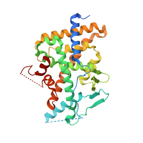
    6DUP

  • PubMed Abstract: 

    This work describes the rational amelioration of Cytochrome P450 4/5 (CYP3A4/5) induction through the Pregnane-X Receptor (PXR) pathway in a series of compounds that modulate the metabotropic glutamate Receptor 2 (mGluR2) via an allosteric mechanism. The compounds were initially shown to induce CYP3A4/5 via the gold-standard induction assay measured in primary human hepatocytes. This was followed up by testing the compounds in a PXR assay which correlated well with the assay in primary cells. Further, one of the compounds was crystallized with PXR (pdb code 6DUP). Analysis of this co-crystal structure, together with previously published PXR co-crystal structures, lead to modification ideas. The compounds synthesized based on these ideas were shown not to be CYP3A4/5 inducers. The mGluR2 activity of the resulting compounds was maintained.


  • Organizational Affiliation

    Integrated Drug Discovery, Sanofi US, 153-2nd Ave., Waltham, MA 02451, USA. Electronic address: roy.vaz@sanofi.com.


Macromolecules
Find similar proteins by:  (by identity cutoff)  |  3D Structure
Entity ID: 1
MoleculeChains Sequence LengthOrganismDetailsImage
Nuclear receptor subfamily 1 group I member 2
A, B
344Homo sapiensMutation(s): 0 
Gene Names: NR1I2PXR
UniProt & NIH Common Fund Data Resources
Find proteins for O75469 (Homo sapiens)
Explore O75469 
Go to UniProtKB:  O75469
PHAROS:  O75469
GTEx:  ENSG00000144852 
Entity Groups  
Sequence Clusters30% Identity50% Identity70% Identity90% Identity95% Identity100% Identity
UniProt GroupO75469
Sequence Annotations
Expand
  • Reference Sequence
Small Molecules
Ligands 1 Unique
IDChains Name / Formula / InChI Key2D Diagram3D Interactions
HCJ
Query on HCJ

Download Ideal Coordinates CCD File 
C [auth A](2S)-2-({[3'-(trifluoromethyl)[1,1'-biphenyl]-4-yl]oxy}methyl)-2,3-dihydro-7H-[1,3]oxazolo[3,2-a]pyrimidin-7-one
C20 H15 F3 N2 O3
AYYWUDDPGRDIQB-KRWDZBQOSA-N
Experimental Data & Validation

Experimental Data

  • Method: X-RAY DIFFRACTION
  • Resolution: 2.30 Å
  • R-Value Free: 0.266 
  • R-Value Work: 0.226 
  • Space Group: P 21 21 21
Unit Cell:
Length ( Å )Angle ( ˚ )
a = 84.034α = 90
b = 88.926β = 90
c = 106.452γ = 90
Software Package:
Software NamePurpose
CNXrefinement
HKL-2000data collection
SCALEPACKdata scaling
PHASERphasing
HKL-2000data reduction

Structure Validation

View Full Validation Report


Ligand Structure Quality Assessment 


Entry History 

Deposition Data

Revision History  (Full details and data files)

  • Version 1.0: 2018-08-29
    Type: Initial release
  • Version 1.1: 2018-09-12
    Changes: Data collection, Database references
  • Version 1.2: 2018-10-17
    Changes: Data collection, Database references
  • Version 1.3: 2024-03-13
    Changes: Data collection, Database references