6DJU

Mtb ClpB in complex with ATPgammaS and casein, Conformer 1


Experimental Data Snapshot

  • Method: ELECTRON MICROSCOPY
  • Resolution: 3.80 Å
  • Aggregation State: PARTICLE 
  • Reconstruction Method: SINGLE PARTICLE 

wwPDB Validation   3D Report Full Report


This is version 1.4 of the entry. See complete history


Literature

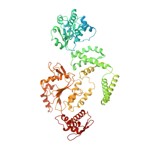
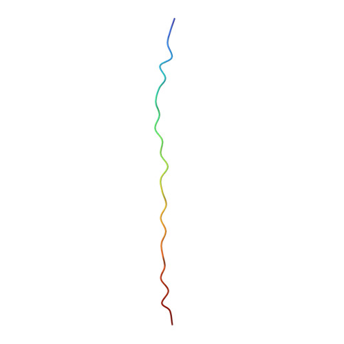
ATP hydrolysis-coupled peptide translocation mechanism ofMycobacterium tuberculosisClpB.

Yu, H.Lupoli, T.J.Kovach, A.Meng, X.Zhao, G.Nathan, C.F.Li, H.

(2018) Proc Natl Acad Sci U S A 115: E9560-E9569

  • DOI: https://doi.org/10.1073/pnas.1810648115
  • Primary Citation of Related Structures:  
    6DJU, 6DJV, 6ED3

  • PubMed Abstract: 

    The protein disaggregase ClpB hexamer is conserved across evolution and has two AAA+-type nucleotide-binding domains, NBD1 and NBD2, in each protomer. In M. tuberculosis ( Mtb ), ClpB facilitates asymmetric distribution of protein aggregates during cell division to help the pathogen survive and persist within the host, but a mechanistic understanding has been lacking. Here we report cryo-EM structures at 3.8- to 3.9-Å resolution of Mtb ClpB bound to a model substrate, casein, in the presence of the weakly hydrolyzable ATP mimic adenosine 5'-[γ-thio]triphosphate. Mtb ClpB existed in solution in two closed-ring conformations, conformers 1 and 2. In both conformers, the 12 pore-loops on the 12 NTDs of the six protomers (P1-P6) were arranged similarly to a staircase around the bound peptide. Conformer 1 is a low-affinity state in which three of the 12 pore-loops (the protomer P1 NBD1 and NBD2 loops and the protomer P2 NBD1 loop) are not engaged with peptide. Conformer 2 is a high-affinity state because only one pore-loop (the protomer P2 NBD1 loop) is not engaged with the peptide. The resolution of the two conformations, along with their bound substrate peptides and nucleotides, enabled us to propose a nucleotide-driven peptide translocation mechanism of a bacterial ClpB that is largely consistent with several recent unfoldase structures, in particular with the eukaryotic Hsp104. However, whereas Hsp104's two NBDs move in opposing directions during one step of peptide translocation, in Mtb ClpB the two NBDs move only in the direction of translocation.


  • Organizational Affiliation

    Cryo-Electron Microscopy Structural Biology Laboratory, Van Andel Research Institute, Grand Rapids, MI 49503.


Macromolecules
Find similar proteins by:  (by identity cutoff)  |  3D Structure
Entity ID: 1
MoleculeChains Sequence LengthOrganismDetailsImage
Chaperone protein ClpB848Mycobacterium tuberculosisMutation(s): 0 
Gene Names: clpBclpB_1ERS023446_03763ERS124361_00048SAMEA2682864_01140SAMEA2683035_02692
UniProt
Find proteins for P9WPD1 (Mycobacterium tuberculosis (strain ATCC 25618 / H37Rv))
Explore P9WPD1 
Go to UniProtKB:  P9WPD1
Entity Groups  
Sequence Clusters30% Identity50% Identity70% Identity90% Identity95% Identity100% Identity
UniProt GroupP9WPD1
Sequence Annotations
Expand
  • Reference Sequence
Find similar proteins by:  (by identity cutoff)  |  3D Structure
Entity ID: 2
MoleculeChains Sequence LengthOrganismDetailsImage
casein polyAlanine modelE [auth N]26Bos taurusMutation(s): 0 
Entity Groups  
Sequence Clusters30% Identity50% Identity70% Identity90% Identity95% Identity100% Identity
Sequence Annotations
Expand
  • Reference Sequence
Small Molecules
Ligands 2 Unique
IDChains Name / Formula / InChI Key2D Diagram3D Interactions
AGS
Query on AGS

Download Ideal Coordinates CCD File 
H [auth A]
I [auth A]
J [auth B]
K [auth B]
L [auth C]
H [auth A],
I [auth A],
J [auth B],
K [auth B],
L [auth C],
M [auth C],
N [auth D],
O [auth D],
P [auth E],
R [auth F]
PHOSPHOTHIOPHOSPHORIC ACID-ADENYLATE ESTER
C10 H16 N5 O12 P3 S
NLTUCYMLOPLUHL-KQYNXXCUSA-N
ADP
Query on ADP

Download Ideal Coordinates CCD File 
Q [auth E],
S [auth F]
ADENOSINE-5'-DIPHOSPHATE
C10 H15 N5 O10 P2
XTWYTFMLZFPYCI-KQYNXXCUSA-N
Experimental Data & Validation

Experimental Data

  • Method: ELECTRON MICROSCOPY
  • Resolution: 3.80 Å
  • Aggregation State: PARTICLE 
  • Reconstruction Method: SINGLE PARTICLE 
EM Software:
TaskSoftware PackageVersion
RECONSTRUCTIONRELION2.0

Structure Validation

View Full Validation Report


Entry History 

Deposition Data

Revision History  (Full details and data files)

  • Version 1.0: 2018-09-26
    Type: Initial release
  • Version 1.1: 2018-10-10
    Changes: Data collection, Database references
  • Version 1.2: 2018-10-24
    Changes: Data collection, Database references
  • Version 1.3: 2019-12-18
    Changes: Other
  • Version 1.4: 2024-03-13
    Changes: Data collection, Database references