6CNY

2.3 Angstrom Structure of Phosphodiesterase treated Vivid (complex with FMN)


Experimental Data Snapshot

  • Method: X-RAY DIFFRACTION
  • Resolution: 2.10 Å
  • R-Value Free: 0.204 
  • R-Value Work: 0.163 
  • R-Value Observed: 0.164 

wwPDB Validation   3D Report Full Report


Ligand Structure Quality Assessment 


This is version 1.4 of the entry. See complete history


Literature

Conformational switching in the fungal light sensor Vivid.

Zoltowski, B.D.Schwerdtfeger, C.Widom, J.Loros, J.J.Bilwes, A.M.Dunlap, J.C.Crane, B.R.

(2007) Science 316: 1054-1057

  • DOI: https://doi.org/10.1126/science.1137128
  • Primary Citation of Related Structures:  
    2PD7, 2PD8, 2PDR, 6CNY

  • PubMed Abstract: 

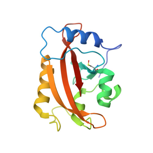
    The Neurospora crassa photoreceptor Vivid tunes blue-light responses and modulates gating of the circadian clock. Crystal structures of dark-state and light-state Vivid reveal a light, oxygen, or voltage Per-Arnt-Sim domain with an unusual N-terminal cap region and a loop insertion that accommodates the flavin cofactor. Photoinduced formation of a cystein-flavin adduct drives flavin protonation to induce an N-terminal conformational change. A cysteine-to-serine substitution remote from the flavin adenine dinucleotide binding site decouples conformational switching from the flavin photocycle and prevents Vivid from sending signals in Neurospora. Key elements of this activation mechanism are conserved by other photosensors such as White Collar-1, ZEITLUPE, ENVOY, and flavin-binding, kelch repeat, F-BOX 1 (FKF1).


  • Organizational Affiliation

    Department of Chemistry and Chemical Biology, Cornell University, Ithaca, NY 14853, USA.


Macromolecules
Find similar proteins by:  (by identity cutoff)  |  3D Structure
Entity ID: 1
MoleculeChains Sequence LengthOrganismDetailsImage
Vivid PAS protein VVD
A, B, C, D
151Neurospora crassaMutation(s): 0 
Gene Names: vvdG17A4.050GE21DRAFT_9175
UniProt
Find proteins for Q9C3Y6 (Neurospora crassa)
Explore Q9C3Y6 
Go to UniProtKB:  Q9C3Y6
Entity Groups  
Sequence Clusters30% Identity50% Identity70% Identity90% Identity95% Identity100% Identity
UniProt GroupQ9C3Y6
Sequence Annotations
Expand
  • Reference Sequence
Experimental Data & Validation

Experimental Data

  • Method: X-RAY DIFFRACTION
  • Resolution: 2.10 Å
  • R-Value Free: 0.204 
  • R-Value Work: 0.163 
  • R-Value Observed: 0.164 
  • Space Group: P 1 21 1
Unit Cell:
Length ( Å )Angle ( ˚ )
a = 64.12α = 90
b = 80.57β = 90.02
c = 64.11γ = 90
Software Package:
Software NamePurpose
REFMACrefinement
PDB_EXTRACTdata extraction
HKL-2000data collection
HKL-2000data reduction
HKL-2000data scaling
AMoREphasing

Structure Validation

View Full Validation Report


Ligand Structure Quality Assessment 


Entry History & Funding Information

Deposition Data


Funding OrganizationLocationGrant Number
National Institutes of Health/National Institute of General Medical Sciences (NIH/NIGMS)United States--
Searle Scholars ProgramUnited States--

Revision History  (Full details and data files)

  • Version 1.0: 2018-03-21
    Type: Initial release
  • Version 1.1: 2018-10-10
    Changes: Data collection, Database references, Structure summary
  • Version 1.2: 2019-02-20
    Changes: Author supporting evidence, Data collection, Structure summary
  • Version 1.3: 2022-03-23
    Changes: Author supporting evidence, Database references
  • Version 1.4: 2023-10-04
    Changes: Data collection, Refinement description