6BP2

Therapeutic human monoclonal antibody MR191 bound to a marburgvirus glycoprotein


Experimental Data Snapshot

  • Method: X-RAY DIFFRACTION
  • Resolution: 3.17 Å
  • R-Value Free: 0.284 
  • R-Value Work: 0.252 
  • R-Value Observed: 0.254 

wwPDB Validation   3D Report Full Report


This is version 3.1 of the entry. See complete history


Literature

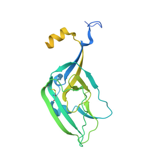
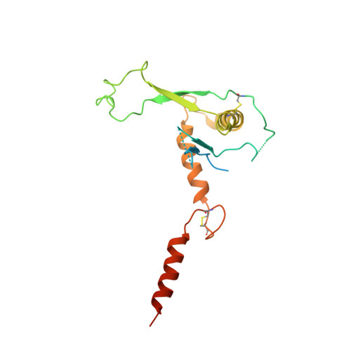
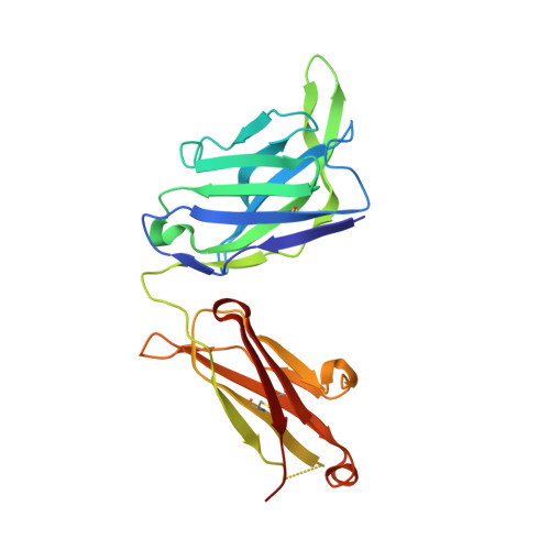
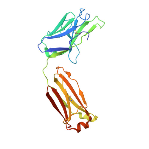
The Marburgvirus-Neutralizing Human Monoclonal Antibody MR191 Targets a Conserved Site to Block Virus Receptor Binding.

King, L.B.Fusco, M.L.Flyak, A.I.Ilinykh, P.A.Huang, K.Gunn, B.Kirchdoerfer, R.N.Hastie, K.M.Sangha, A.K.Meiler, J.Alter, G.Bukreyev, A.Crowe, J.E.Saphire, E.O.

(2018) Cell Host Microbe 23: 101-109.e4

  • DOI: https://doi.org/10.1016/j.chom.2017.12.003
  • Primary Citation of Related Structures:  
    6BP2

  • PubMed Abstract: 

    Since their first identification 50 years ago, marburgviruses have emerged several times, with 83%-90% lethality in the largest outbreaks. Although no vaccines or therapeutics are available for human use, the human antibody MR191 provides complete protection in non-human primates when delivered several days after inoculation of a lethal marburgvirus dose. The detailed neutralization mechanism of MR191 remains outstanding. Here we present a 3.2 Å crystal structure of MR191 complexed with a trimeric marburgvirus surface glycoprotein (GP). MR191 neutralizes by occupying the conserved receptor-binding site and competing with the host receptor Niemann-Pick C1. The structure illuminates previously disordered regions of GP including the stalk, fusion loop, CX 6 CC switch, and an N-terminal region of GP2 that wraps about the outside of GP1 to anchor a marburgvirus-specific "wing" antibody epitope. Virus escape mutations mapped far outside the MR191 receptor-binding site footprint suggest a role for these other regions in the GP quaternary structure.


  • Organizational Affiliation

    Department of Immunology and Microbiology, The Scripps Research Institute, La Jolla, CA 92037, USA.


Macromolecules
Find similar proteins by:  (by identity cutoff)  |  3D Structure
Entity ID: 1
MoleculeChains Sequence LengthOrganismDetailsImage
Envelope glycoprotein250Orthomarburgvirus marburgenseMutation(s): 9 
Gene Names: GPDF49_53427gpGP
UniProt
Find proteins for Q1PDC7 (Lake Victoria marburgvirus (strain Ravn-87))
Explore Q1PDC7 
Go to UniProtKB:  Q1PDC7
Entity Groups  
Sequence Clusters30% Identity50% Identity70% Identity90% Identity95% Identity100% Identity
UniProt GroupQ1PDC7
Sequence Annotations
Expand
  • Reference Sequence
Find similar proteins by:  (by identity cutoff)  |  3D Structure
Entity ID: 2
MoleculeChains Sequence LengthOrganismDetailsImage
Envelope glycoprotein GP2202Orthomarburgvirus marburgenseMutation(s): 4 
Gene Names: GPDF49_53427gpGP
UniProt
Find proteins for Q1PDC7 (Lake Victoria marburgvirus (strain Ravn-87))
Explore Q1PDC7 
Go to UniProtKB:  Q1PDC7
Entity Groups  
Sequence Clusters30% Identity50% Identity70% Identity90% Identity95% Identity100% Identity
UniProt GroupQ1PDC7
Sequence Annotations
Expand
  • Reference Sequence
Find similar proteins by:  (by identity cutoff)  |  3D Structure
Entity ID: 3
MoleculeChains Sequence LengthOrganismDetailsImage
MR191 Fab Heavy ChainC [auth H]229Homo sapiensMutation(s): 0 
Entity Groups  
Sequence Clusters30% Identity50% Identity70% Identity90% Identity95% Identity100% Identity
Sequence Annotations
Expand
  • Reference Sequence
Find similar proteins by:  (by identity cutoff)  |  3D Structure
Entity ID: 4
MoleculeChains Sequence LengthOrganismDetailsImage
MR191 Fab Light ChainD [auth L]217Homo sapiensMutation(s): 0 
Entity Groups  
Sequence Clusters30% Identity50% Identity70% Identity90% Identity95% Identity100% Identity
Sequence Annotations
Expand
  • Reference Sequence
Oligosaccharides

Help

Entity ID: 5
MoleculeChains Length2D Diagram Glycosylation3D Interactions
beta-D-mannopyranose-(1-4)-2-acetamido-2-deoxy-beta-D-glucopyranose-(1-4)-2-acetamido-2-deoxy-beta-D-glucopyranoseE [auth C]3N-Glycosylation
Glycosylation Resources
GlyTouCan:  G15407YE
GlyCosmos:  G15407YE
GlyGen:  G15407YE
Entity ID: 6
MoleculeChains Length2D Diagram Glycosylation3D Interactions
2-acetamido-2-deoxy-beta-D-glucopyranose-(1-4)-2-acetamido-2-deoxy-beta-D-glucopyranoseF [auth D],
H [auth F]
2N-Glycosylation
Glycosylation Resources
GlyTouCan:  G42666HT
GlyCosmos:  G42666HT
GlyGen:  G42666HT
Entity ID: 7
MoleculeChains Length2D Diagram Glycosylation3D Interactions
alpha-D-mannopyranose-(1-3)-[alpha-D-mannopyranose-(1-6)]beta-D-mannopyranoseG [auth E]3N/A
Glycosylation Resources
GlyTouCan:  G72618IM
GlyCosmos:  G72618IM
GlyGen:  G72618IM
Experimental Data & Validation

Experimental Data

  • Method: X-RAY DIFFRACTION
  • Resolution: 3.17 Å
  • R-Value Free: 0.284 
  • R-Value Work: 0.252 
  • R-Value Observed: 0.254 
  • Space Group: P 3 2 1
Unit Cell:
Length ( Å )Angle ( ˚ )
a = 133.796α = 90
b = 133.796β = 90
c = 151.003γ = 120
Software Package:
Software NamePurpose
PHENIXrefinement
XDSdata reduction
XDSdata scaling
PHENIXphasing

Structure Validation

View Full Validation Report


Entry History & Funding Information

Deposition Data


Funding OrganizationLocationGrant Number
National Institutes of Health/National Institute Of Allergy and Infectious Diseases (NIH/NIAID)United StatesR01AI089498
National Institutes of Health/National Institute Of Allergy and Infectious Diseases (NIH/NIAID)United StatesU19AI109762

Revision History  (Full details and data files)

  • Version 1.0: 2018-01-17
    Type: Initial release
  • Version 1.1: 2018-01-31
    Changes: Database references
  • Version 2.0: 2018-03-07
    Changes: Advisory, Atomic model, Derived calculations
  • Version 2.1: 2019-12-11
    Changes: Author supporting evidence
  • Version 3.0: 2020-07-29
    Type: Remediation
    Reason: Carbohydrate remediation
    Changes: Advisory, Atomic model, Data collection, Derived calculations, Structure summary
  • Version 3.1: 2023-10-04
    Changes: Data collection, Database references, Derived calculations, Refinement description, Structure summary