6APX

Crystal structure of human dual specificity phosphatase 1 catalytic domain (C258S) as a maltose binding protein fusion in complex with the monobody YSX1


Experimental Data Snapshot

  • Method: X-RAY DIFFRACTION
  • Resolution: 2.49 Å
  • R-Value Free: 0.236 
  • R-Value Work: 0.207 
  • R-Value Observed: 0.208 

wwPDB Validation   3D Report Full Report


This is version 1.3 of the entry. See complete history


Literature

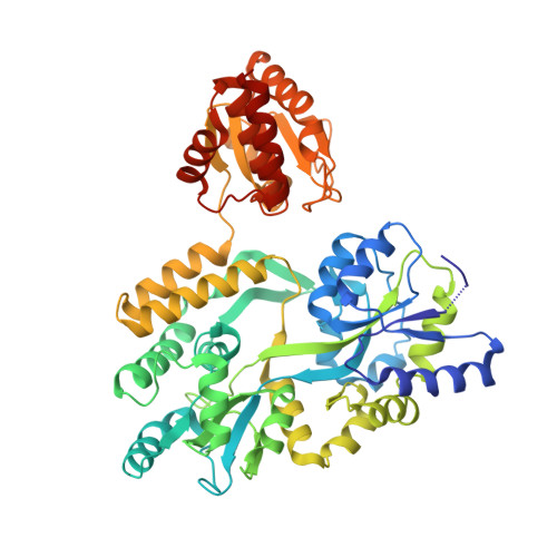
Crystal structure of the human dual specificity phosphatase 1 catalytic domain.

Gumpena, R.Lountos, G.T.Raran-Kurussi, S.Tropea, J.E.Cherry, S.Waugh, D.S.

(2018) Protein Sci 27: 561-567

  • DOI: https://doi.org/10.1002/pro.3328
  • Primary Citation of Related Structures:  
    6APX

  • PubMed Abstract: 

    The dual specificity phosphatase DUSP1 was the first mitogen activated protein kinase phosphatase (MKP) to be identified. It dephosphorylates conserved tyrosine and threonine residues in the activation loops of mitogen activated protein kinases ERK2, JNK1 and p38-alpha. Here, we report the crystal structure of the human DUSP1 catalytic domain at 2.49 Å resolution. Uniquely, the protein was crystallized as an MBP fusion protein in complex with a monobody that binds to MBP. Sulfate ions occupy the phosphotyrosine and putative phosphothreonine binding sites in the DUSP1 catalytic domain.


  • Organizational Affiliation

    Macromolecular Crystallography Laboratory, Center for Cancer Research, National Cancer Institute at Frederick, Frederick, MD, 21702.


Macromolecules
Find similar proteins by:  (by identity cutoff)  |  3D Structure
Entity ID: 1
MoleculeChains Sequence LengthOrganismDetailsImage
Maltose-binding periplasmic protein,Dual specificity protein phosphatase 1520Escherichia coli K-12Homo sapiens
This entity is chimeric
Mutation(s): 9 
Gene Names: malEb4034JW3994DUSP1CL100MKP1PTPN10VH1
EC: 3.1.3.16 (PDB Primary Data), 3.1.3.48 (PDB Primary Data)
UniProt & NIH Common Fund Data Resources
Find proteins for P0AEX9 (Escherichia coli (strain K12))
Explore P0AEX9 
Go to UniProtKB:  P0AEX9
Find proteins for P28562 (Homo sapiens)
Explore P28562 
Go to UniProtKB:  P28562
PHAROS:  P28562
GTEx:  ENSG00000120129 
Entity Groups  
Sequence Clusters30% Identity50% Identity70% Identity90% Identity95% Identity100% Identity
UniProt GroupsP28562P0AEX9
Sequence Annotations
Expand
  • Reference Sequence
Find similar proteins by:  (by identity cutoff)  |  3D Structure
Entity ID: 2
MoleculeChains Sequence LengthOrganismDetailsImage
Monobody YSX196synthetic constructMutation(s): 0 
Entity Groups  
Sequence Clusters30% Identity50% Identity70% Identity90% Identity95% Identity100% Identity
Sequence Annotations
Expand
  • Reference Sequence
Experimental Data & Validation

Experimental Data

  • Method: X-RAY DIFFRACTION
  • Resolution: 2.49 Å
  • R-Value Free: 0.236 
  • R-Value Work: 0.207 
  • R-Value Observed: 0.208 
  • Space Group: P 43 21 2
Unit Cell:
Length ( Å )Angle ( ˚ )
a = 105.802α = 90
b = 105.802β = 90
c = 181.463γ = 90
Software Package:
Software NamePurpose
PHENIXrefinement
HKL-3000data reduction
HKL-3000data scaling
MOLREPphasing

Structure Validation

View Full Validation Report


Entry History 

Deposition Data

Revision History  (Full details and data files)

  • Version 1.0: 2017-11-01
    Type: Initial release
  • Version 1.1: 2018-01-31
    Changes: Database references
  • Version 1.2: 2018-05-02
    Changes: Data collection, Structure summary
  • Version 1.3: 2023-10-04
    Changes: Data collection, Database references, Refinement description