6AKL

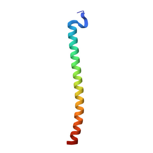
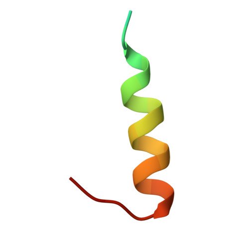
Crystal structure of Striatin3 in complex with SIKE1 Coiled-coil domain


Experimental Data Snapshot

  • Method: X-RAY DIFFRACTION
  • Resolution: 1.75 Å
  • R-Value Free: 0.213 
  • R-Value Work: 0.187 
  • R-Value Observed: 0.189 

wwPDB Validation   3D Report Full Report


This is version 1.2 of the entry. See complete history


Literature

Architecture, substructures, and dynamic assembly of STRIPAK complexes in Hippo signaling.

Tang, Y.Chen, M.Zhou, L.Ma, J.Li, Y.Zhang, H.Shi, Z.Xu, Q.Zhang, X.Gao, Z.Zhao, Y.Cheng, Y.Jiao, S.Zhou, Z.

(2019) Cell Discov 5: 3-3

  • DOI: https://doi.org/10.1038/s41421-018-0077-3
  • Primary Citation of Related Structures:  
    6AKK, 6AKL, 6AKM

  • PubMed Abstract: 

    Striatin-interacting phosphatases and kinases (STRIPAKs) are evolutionarily conserved supramolecular complexes, which have been implicated in the Hippo signaling pathway. Yet the topological structure and dynamic assembly of STRIPAK complexes remain elusive. Here, we report the overall architecture and substructures of a Hippo kinase-containing STRIPAK complex. PP2Aa/c-bound STRN3 directly contacts the Hippo kinase MST2 and also controls the loading of MST2 via two "arms" in a phosphorylation-dependent manner, one arm being STRIP1 and the other SIKE1-SLMAP. A decreased cell density triggered the dissociation of the STRIP1 arm from STRIPAK, reflecting the dynamic assembly of the complex upon sensing upstream signals. Crystallographic studies defined at atomic resolution the interface between STRN3 and SIKE1, and that between SIKE1 and SLMAP. Disrupting the complex assembly abrogated the regulatory effect of STRIPAK towards Hippo signaling. Collectively, our study revealed a "two-arm" assembly of STRIPAK with context-dependent dynamics, offering a framework for further studies on Hippo signaling and biological processes involving MST kinases.


  • Organizational Affiliation

    1State Key Laboratory of Cell Biology, CAS Center for Excellence in Molecular Cell Science, Shanghai Institute of Biochemistry and Cell Biology, Chinese Academy of Sciences, University of Chinese Academy of Sciences, Shanghai, 200031 China.


Macromolecules
Find similar proteins by:  (by identity cutoff)  |  3D Structure
Entity ID: 1
MoleculeChains Sequence LengthOrganismDetailsImage
Suppressor of IKBKE 1
A, B
54Homo sapiensMutation(s): 0 
Gene Names: SIKE1SIKE
UniProt & NIH Common Fund Data Resources
Find proteins for Q9BRV8 (Homo sapiens)
Explore Q9BRV8 
Go to UniProtKB:  Q9BRV8
PHAROS:  Q9BRV8
GTEx:  ENSG00000052723 
Entity Groups  
Sequence Clusters30% Identity50% Identity70% Identity90% Identity95% Identity100% Identity
UniProt GroupQ9BRV8
Sequence Annotations
Expand
  • Reference Sequence
Find similar proteins by:  (by identity cutoff)  |  3D Structure
Entity ID: 2
MoleculeChains Sequence LengthOrganismDetailsImage
Striatin-326Homo sapiensMutation(s): 0 
UniProt & NIH Common Fund Data Resources
Find proteins for Q13033 (Homo sapiens)
Explore Q13033 
Go to UniProtKB:  Q13033
PHAROS:  Q13033
GTEx:  ENSG00000196792 
Entity Groups  
Sequence Clusters30% Identity50% Identity70% Identity90% Identity95% Identity100% Identity
UniProt GroupQ13033
Sequence Annotations
Expand
  • Reference Sequence
Experimental Data & Validation

Experimental Data

  • Method: X-RAY DIFFRACTION
  • Resolution: 1.75 Å
  • R-Value Free: 0.213 
  • R-Value Work: 0.187 
  • R-Value Observed: 0.189 
  • Space Group: P 21 21 21
Unit Cell:
Length ( Å )Angle ( ˚ )
a = 39.282α = 90
b = 42.382β = 90
c = 91.228γ = 90
Software Package:
Software NamePurpose
PHENIXrefinement
HKL-3000data reduction
HKL-3000data scaling
PHENIXphasing

Structure Validation

View Full Validation Report


Entry History 

Deposition Data

Revision History  (Full details and data files)

  • Version 1.0: 2019-01-16
    Type: Initial release
  • Version 1.1: 2019-02-13
    Changes: Data collection, Database references
  • Version 1.2: 2023-11-22
    Changes: Data collection, Database references, Refinement description