


Structural basis for nucleotide-mediated remodelling mechanism of Mycobacterium Mfd
Putta, S., Prabha, S., Bhat, V., Fox, G.C., Walsh, M.A., Rao, D.N., Nagaraja, V., Natesh, R.To be published.
Experimental Data Snapshot
Entity ID: 1 | |||||
|---|---|---|---|---|---|
| Molecule | Chains | Sequence Length | Organism | Details | Image |
| Mycobacterium smegmatis Mfd | 1,235 | Mycolicibacterium smegmatis MC2 155 | Mutation(s): 0 Gene Names: mfd EC: 3.6.4 | 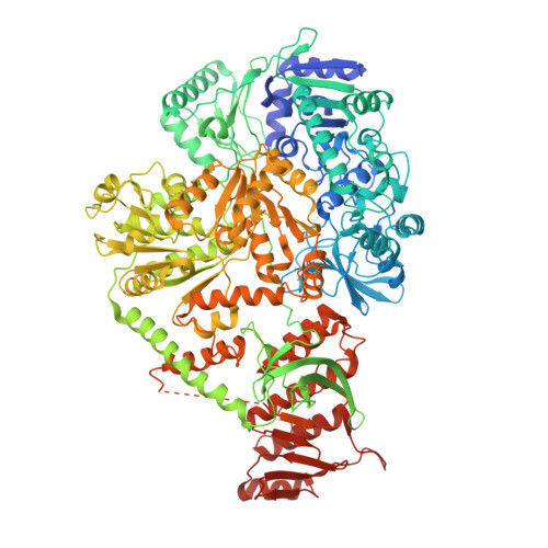 | |
UniProt | |||||
Find proteins for A0R3C5 (Mycolicibacterium smegmatis (strain ATCC 700084 / mc(2)155)) Explore A0R3C5 Go to UniProtKB: A0R3C5 | |||||
Entity Groups | |||||
| Sequence Clusters | 30% Identity50% Identity70% Identity90% Identity95% Identity100% Identity | ||||
| UniProt Group | A0R3C5 | ||||
Sequence AnnotationsExpand | |||||
| |||||
| Ligands 3 Unique | |||||
|---|---|---|---|---|---|
| ID | Chains | Name / Formula / InChI Key | 2D Diagram | 3D Interactions | |
| ADP Query on ADP | G [auth A], I [auth B] | ADENOSINE-5'-DIPHOSPHATE C10 H15 N5 O10 P2 XTWYTFMLZFPYCI-KQYNXXCUSA-N |  | ||
| SO4 Query on SO4 | C [auth A], D [auth A], E [auth A], H [auth B] | SULFATE ION O4 S QAOWNCQODCNURD-UHFFFAOYSA-L |  | ||
| PO4 Query on PO4 | F [auth A] | PHOSPHATE ION O4 P NBIIXXVUZAFLBC-UHFFFAOYSA-K |  | ||
| Length ( Å ) | Angle ( ˚ ) |
|---|---|
| a = 83.659 | α = 90 |
| b = 160.44 | β = 90 |
| c = 214.529 | γ = 90 |
| Software Name | Purpose |
|---|---|
| PHENIX | refinement |
| iMOSFLM | data reduction |
| SCALA | data scaling |
| MOLREP | phasing |
| Funding Organization | Location | Grant Number |
|---|---|---|
| Other government | India | BT/HRD/35/02/19/2009 |
RCSB PDB (citation) is hosted by
RCSB PDB is a member of the

