6VU6

Sialic acid binding region of Streptococcus Sanguinis SK1 adhesin bound to 3'sLn

  • Classification: CELL ADHESION
  • Organism(s): Streptococcus sanguinis SK1 = NCTC 7863
  • Expression System: Escherichia coli
  • Mutation(s): No 

  • Deposited: 2020-02-14 Released: 2020-08-26 
  • Deposition Author(s): Stubbs, H.E., Iverson, T.M.
  • Funding Organization(s): National Institutes of Health/National Institute of General Medical Sciences (NIH/NIGMS), National Institutes of Health/National Eye Institute (NIH/NEI), National Institutes of Health/National Institute Of Allergy and Infectious Diseases (NIH/NIAID), National Institutes of Health/National Institute of Dental and Craniofacial Research (NIH/NIDCR)

Experimental Data Snapshot

  • Method: X-RAY DIFFRACTION
  • Resolution: 2.10 Å
  • R-Value Free: 0.248 
  • R-Value Work: 0.225 
  • R-Value Observed: 0.226 

wwPDB Validation   3D Report Full Report

Currently 6VU6 does not have a validation slider image.


This is version 1.2 of the entry. See complete history


Literature

Tandem sialoglycan-binding modules in a Streptococcus sanguinis serine-rich repeat adhesin create target dependent avidity effects.

Stubbs, H.E.Bensing, B.A.Yamakawa, I.Sharma, P.Yu, H.Chen, X.Sullam, P.M.Iverson, T.M.

(2020) J Biol Chem 295: 14737-14749

  • DOI: https://doi.org/10.1074/jbc.RA120.014177
  • Primary Citation of Related Structures:  
    6VS7, 6VT2, 6VU6

  • PubMed Abstract: 

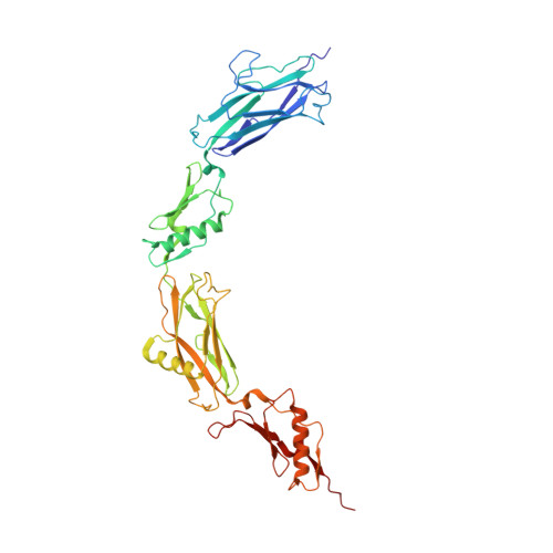
    S ialic acid-binding i mmuno g lobulin-like lec tins (Siglec)-like domains of streptococcal serine-rich repeat (SRR) adhesins recognize sialylated glycans on human salivary, platelet, and plasma glycoproteins via a YTRY sequence motif. The SRR adhesin from Streptococcus sanguinis strain SK1 has tandem sialoglycan-binding domains and has previously been shown to bind sialoglycans with high affinity. However, both domains contain substitutions within the canonical YTRY motif, making it unclear how they interact with host receptors. To identify how the S. sanguinis strain SK1 SRR adhesin affects interactions with sialylated glycans and glycoproteins, we determined high-resolution crystal structures of the binding domains alone and with purified trisaccharides. These structural studies determined that the ligands still bind at the noncanonical binding motif, but with fewer hydrogen-bonding interactions to the protein than is observed in structures of other Siglec-like adhesins. Complementary biochemical studies identified that each of the two binding domains has a different selectivity profile. Interestingly, the binding of SK1 to platelets and plasma glycoproteins identified that the interaction to some host targets is dominated by the contribution of one binding domain, whereas the binding to other host receptors is mediated by both binding domains. These results provide insight into outstanding questions concerning the roles of tandem domains in targeting host receptors and suggest mechanisms for how pathogens can adapt to the availability of a range of related but nonidentical host receptors. They further suggest that the definition of the YTRY motif should be changed to ϕTR X , a more rigorous description of this sialic acid-recognition motif given recent findings.


  • Organizational Affiliation

    Graduate Program in Chemical and Physical Biology, Vanderbilt University, Nashville, Tennessee, USA.


Macromolecules
Find similar proteins by:  (by identity cutoff)  |  3D Structure
Entity ID: 1
MoleculeChains Sequence LengthOrganismDetailsImage
AdhesinA,
B [auth E]
409Streptococcus sanguinis SK1 = NCTC 7863Mutation(s): 0 
Entity Groups  
Sequence Clusters30% Identity50% Identity70% Identity90% Identity95% Identity100% Identity
Sequence Annotations
Expand
  • Reference Sequence
Oligosaccharides

Help

Entity ID: 2
MoleculeChains Length2D Diagram Glycosylation3D Interactions
N-acetyl-alpha-neuraminic acid-(2-3)-beta-D-galactopyranose-(1-4)-2-acetamido-2-deoxy-beta-D-glucopyranoseC [auth B],
D [auth C],
E [auth D],
F
3N/A
Glycosylation Resources
GlyTouCan:  G00065MO
GlyCosmos:  G00065MO
GlyGen:  G00065MO
Experimental Data & Validation

Experimental Data

Unit Cell:
Length ( Å )Angle ( ˚ )
a = 82.213α = 90
b = 269.859β = 90
c = 47.511γ = 90
Software Package:
Software NamePurpose
PHENIXrefinement
HKL-2000data reduction
HKL-2000data scaling
PHASERphasing

Structure Validation

View Full Validation Report

Currently 6VU6 does not have a validation slider image.


Entry History & Funding Information

Deposition Data


Funding OrganizationLocationGrant Number
National Institutes of Health/National Institute of General Medical Sciences (NIH/NIGMS)United States5T32GM008320-28
National Institutes of Health/National Eye Institute (NIH/NEI)United States5T32EY007135-24
National Institutes of Health/National Institute Of Allergy and Infectious Diseases (NIH/NIAID)United StatesAI106987
National Institutes of Health/National Institute Of Allergy and Infectious Diseases (NIH/NIAID)United StatesAI10400
National Institutes of Health/National Institute of Dental and Craniofacial Research (NIH/NIDCR)United StatesDE019807

Revision History  (Full details and data files)

  • Version 1.0: 2020-08-26
    Type: Initial release
  • Version 1.1: 2021-03-10
    Changes: Database references
  • Version 1.2: 2023-10-11
    Changes: Data collection, Database references, Refinement description