6ICT

Structure of SETD3 bound to SAH and methylated actin


Experimental Data Snapshot

  • Method: X-RAY DIFFRACTION
  • Resolution: 1.95 Å
  • R-Value Free: 0.214 
  • R-Value Work: 0.178 
  • R-Value Observed: 0.180 

wwPDB Validation   3D Report Full Report


Ligand Structure Quality Assessment 


This is version 2.1 of the entry. See complete history


Literature

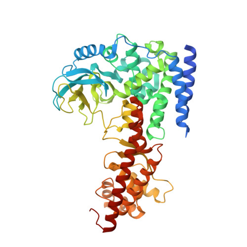
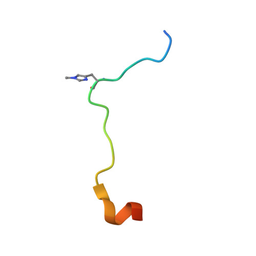
Structural insights into SETD3-mediated histidine methylation on beta-actin.

Guo, Q.Liao, S.Kwiatkowski, S.Tomaka, W.Yu, H.Wu, G.Tu, X.Min, J.Drozak, J.Xu, C.

(2019) Elife 8

  • DOI: https://doi.org/10.7554/eLife.43676
  • Primary Citation of Related Structures:  
    6ICT, 6ICV

  • PubMed Abstract: 

    SETD3 is a member of the SET (Su(var)3-9, Enhancer of zeste, and Trithorax) domain protein superfamily and plays important roles in hypoxic pulmonary hypertension, muscle differentiation, and carcinogenesis. Previously, we identified SETD3 as the actin-specific methyltransferase that methylates the N3 of His73 on β-actin (Kwiatkowski et al., 2018). Here, we present two structures of S -adenosyl-L-homocysteine-bound SETD3 in complex with either an unmodified β-actin peptide or its His-methylated variant. Structural analyses, supported by biochemical experiments and enzyme activity assays, indicate that the recognition and methylation of β-actin by SETD3 are highly sequence specific, and that both SETD3 and β-actin adopt pronounced conformational changes upon binding to each other. In conclusion, this study is the first to show a catalytic mechanism of SETD3-mediated histidine methylation on β-actin, which not only throws light on the protein histidine methylation phenomenon but also facilitates the design of small molecule inhibitors of SETD3.


  • Organizational Affiliation

    Division of Molecular and Cellular Biophysics, Hefei National Laboratory for Physical Sciences at the Microscale, School of Life Sciences, University of Science and Technology of China, Hefei, China.


Macromolecules
Find similar proteins by:  (by identity cutoff)  |  3D Structure
Entity ID: 1
MoleculeChains Sequence LengthOrganismDetailsImage
Histone-lysine N-methyltransferase setd3
A, B, C, D
504Homo sapiensMutation(s): 0 
Gene Names: SETD3C14orf154
EC: 2.1.1.43
UniProt & NIH Common Fund Data Resources
Find proteins for Q86TU7 (Homo sapiens)
Explore Q86TU7 
Go to UniProtKB:  Q86TU7
PHAROS:  Q86TU7
GTEx:  ENSG00000183576 
Entity Groups  
Sequence Clusters30% Identity50% Identity70% Identity90% Identity95% Identity100% Identity
UniProt GroupQ86TU7
Sequence Annotations
Expand
  • Reference Sequence

Find similar proteins by:  Sequence   |   3D Structure  

Entity ID: 2
MoleculeChains Sequence LengthOrganismDetailsImage
Actin, cytoplasmic 1E,
F [auth G],
G [auth H],
H [auth I]
23Homo sapiensMutation(s): 0 
Gene Names: ACTB
UniProt & NIH Common Fund Data Resources
Find proteins for P60709 (Homo sapiens)
Explore P60709 
Go to UniProtKB:  P60709
PHAROS:  P60709
GTEx:  ENSG00000075624 
Entity Groups  
Sequence Clusters30% Identity50% Identity70% Identity90% Identity95% Identity100% Identity
UniProt GroupP60709
Sequence Annotations
Expand
  • Reference Sequence
Small Molecules
Modified Residues  1 Unique
IDChains TypeFormula2D DiagramParent
HIC
Query on HIC
E,
F [auth G],
G [auth H],
H [auth I]
L-PEPTIDE LINKINGC7 H11 N3 O2HIS
Experimental Data & Validation

Experimental Data

  • Method: X-RAY DIFFRACTION
  • Resolution: 1.95 Å
  • R-Value Free: 0.214 
  • R-Value Work: 0.178 
  • R-Value Observed: 0.180 
  • Space Group: P 1 21 1
Unit Cell:
Length ( Å )Angle ( ˚ )
a = 59.976α = 90
b = 176.693β = 93.37
c = 125.892γ = 90
Software Package:
Software NamePurpose
PHENIXrefinement
HKL-3000data reduction
HKL-3000data scaling
PHENIXphasing

Structure Validation

View Full Validation Report


Ligand Structure Quality Assessment 


Entry History & Funding Information

Deposition Data


Funding OrganizationLocationGrant Number
National Natural Science Foundation of ChinaChina31570737
National Natural Science Foundation of ChinaChina31770806

Revision History  (Full details and data files)

  • Version 1.0: 2019-02-27
    Type: Initial release
  • Version 2.0: 2019-03-06
    Changes: Data collection, Database references, Polymer sequence
  • Version 2.1: 2023-11-22
    Changes: Data collection, Database references, Refinement description