5YH2

The structure of DrFam20C1 and hFam20A complex


Experimental Data Snapshot

  • Method: X-RAY DIFFRACTION
  • Resolution: 3.55 Å
  • R-Value Free: 0.298 
  • R-Value Work: 0.245 
  • R-Value Observed: 0.248 

wwPDB Validation   3D Report Full Report


Ligand Structure Quality Assessment 


This is version 1.1 of the entry. See complete history


Literature

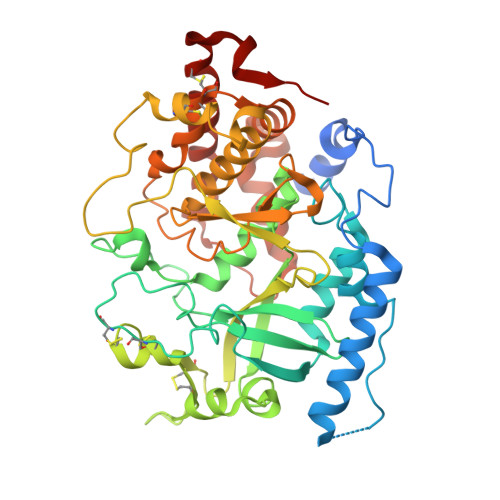
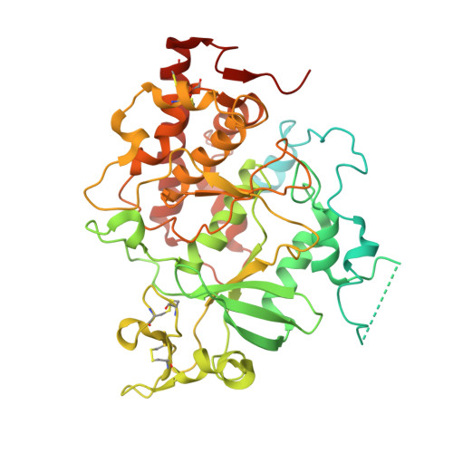
Structure and evolution of the Fam20 kinases

Zhang, H.Zhu, Q.Cui, J.Wang, Y.Chen, M.J.Guo, X.Tagliabracci, V.S.Dixon, J.E.Xiao, J.

(2018) Nat Commun 9: 1218-1218

  • DOI: https://doi.org/10.1038/s41467-018-03615-z
  • Primary Citation of Related Structures:  
    5XOM, 5XOO, 5YH0, 5YH2, 5YH3

  • PubMed Abstract: 

    The Fam20 proteins are novel kinases that phosphorylate secreted proteins and proteoglycans. Fam20C phosphorylates hundreds of secreted proteins and is activated by the pseudokinase Fam20A. Fam20B phosphorylates a xylose residue to regulate proteoglycan synthesis. Despite these wide-ranging and important functions, the molecular and structural basis for the regulation and substrate specificity of these kinases are unknown. Here we report molecular characterizations of all three Fam20 kinases, and show that Fam20C is activated by the formation of an evolutionarily conserved homodimer or heterodimer with Fam20A. Fam20B has a unique active site for recognizing Galβ1-4Xylβ1, the initiator disaccharide within the tetrasaccharide linker region of proteoglycans. We further show that in animals the monomeric Fam20B preceded the appearance of the dimeric Fam20C, and the dimerization trait of Fam20C emerged concomitantly with a change in substrate specificity. Our results provide comprehensive structural, biochemical, and evolutionary insights into the function of the Fam20 kinases.


  • Organizational Affiliation

    The State Key Laboratory of Protein and Plant Gene Research, School of Life Sciences, Peking-Tsinghua Center for Life Sciences, Peking University, 100871, Beijing, China.


Macromolecules
Find similar proteins by:  (by identity cutoff)  |  3D Structure
Entity ID: 1
MoleculeChains Sequence LengthOrganismDetailsImage
Pseudokinase FAM20AA,
C [auth B]
467Homo sapiensMutation(s): 1 
Gene Names: FAM20AUNQ9388/PRO34279
UniProt & NIH Common Fund Data Resources
Find proteins for Q96MK3 (Homo sapiens)
Explore Q96MK3 
Go to UniProtKB:  Q96MK3
PHAROS:  Q96MK3
GTEx:  ENSG00000108950 
Entity Groups  
Sequence Clusters30% Identity50% Identity70% Identity90% Identity95% Identity100% Identity
UniProt GroupQ96MK3
Sequence Annotations
Expand
  • Reference Sequence
Find similar proteins by:  (by identity cutoff)  |  3D Structure
Entity ID: 2
MoleculeChains Sequence LengthOrganismDetailsImage
Family with sequence similarity 20, member CaB [auth C],
D
560Danio rerioMutation(s): 10 
Gene Names: fam20ca
UniProt
Find proteins for E7FBB8 (Danio rerio)
Explore E7FBB8 
Go to UniProtKB:  E7FBB8
Entity Groups  
Sequence Clusters30% Identity50% Identity70% Identity90% Identity95% Identity100% Identity
UniProt GroupE7FBB8
Sequence Annotations
Expand
  • Reference Sequence
Small Molecules
Ligands 1 Unique
IDChains Name / Formula / InChI Key2D Diagram3D Interactions
ATP
Query on ATP

Download Ideal Coordinates CCD File 
E [auth A],
F [auth B]
ADENOSINE-5'-TRIPHOSPHATE
C10 H16 N5 O13 P3
ZKHQWZAMYRWXGA-KQYNXXCUSA-N
Experimental Data & Validation

Experimental Data

  • Method: X-RAY DIFFRACTION
  • Resolution: 3.55 Å
  • R-Value Free: 0.298 
  • R-Value Work: 0.245 
  • R-Value Observed: 0.248 
  • Space Group: P 41 21 2
Unit Cell:
Length ( Å )Angle ( ˚ )
a = 110.059α = 90
b = 110.059β = 90
c = 461.698γ = 90
Software Package:
Software NamePurpose
PHENIXrefinement
HKL-2000data processing
HKL-2000data scaling
PHENIXphasing

Structure Validation

View Full Validation Report


Ligand Structure Quality Assessment 


Entry History 

Deposition Data

Revision History  (Full details and data files)

  • Version 1.0: 2018-04-11
    Type: Initial release
  • Version 1.1: 2018-04-25
    Changes: Data collection, Database references, Source and taxonomy, Structure summary