5YGS

Human TNFRSF25 death domain


Experimental Data Snapshot

  • Method: X-RAY DIFFRACTION
  • Resolution: 2.69 Å
  • R-Value Free: 0.212 
  • R-Value Work: 0.190 

wwPDB Validation   3D Report Full Report


This is version 2.1 of the entry. See complete history


Literature

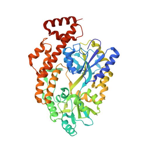
Crystal structure and activation mechanism of DR3 death domain.

Yin, X.Li, W.Ma, H.Zeng, W.Peng, C.Li, Y.Liu, M.Chen, Q.Zhou, R.Jin, T.

(2019) FEBS J 286: 2593-2610

  • DOI: https://doi.org/10.1111/febs.14834
  • Primary Citation of Related Structures:  
    5YEV, 5YGP, 5YGS, 5ZNY, 5ZNZ

  • PubMed Abstract: 

    Death receptor 3 (DR3) (a.k.a. tumor necrosis factor receptor superfamily 25) plays a key role in the immune system by activating nuclear factor kappa-light-chain-enhancer of activated B cells signaling pathway. Here we present the crystal structures of human and mouse DR3 intracellular death domain (DD) at 2.7 and 2.5 Å resolutions, respectively. The mouse DR3 DD adopts a classical six-helix bundle structure while human DR3 DD displays an extended fold. Though there is one-amino-acid difference in the linker between maltose-binding protein (MBP) tag and DR3 DD, according to our self-interaction analysis, the hydrophobic interface discovered in MBP-hDR3 DD crystal structure is responsible for both hDR3 DD and mDR3 DD homotypic interaction. Furthermore, our biochemical analysis indicates that the sequence variation between human and mouse DR3 DD does not affect its structure and function. Small-angle X-ray scattering analysis shows the averaged solution structures of both human and mouse MBP-DR3 DD are the combination of different conformations with different proportion. Through switching to the open conformation, DR3 DD could improve the interaction with downstream element TNFR-associated death domain (TRADD). Here we propose an activation-dependent structural rearrangement model: the DD region is folded as the six-helix bundles in the resting state, while upon extracellular ligand engagement, it switches to the open conformation, which facilitates its self-association and the recruitment of TRADD. Our results provide detailed insights into the architecture of DR3 DD and the molecular mechanism of activation. DATABASES: All refined structure coordinates as well as the corresponding structure factors have been deposited in the PDB under the accession codes 5YGS, 5YEV, 5YGP, 5ZNY, 5ZNZ.


  • Organizational Affiliation

    Hefei National Laboratory for Physical Sciences at Microscale, CAS Key Laboratory of Innate Immunity and Chronic Disease, School of Life Sciences and Medical Center, University of Science and Technology of China, Hefei, Anhui, China.


Macromolecules
Find similar proteins by:  (by identity cutoff)  |  3D Structure
Entity ID: 1
MoleculeChains Sequence LengthOrganismDetailsImage
Human TNRSF25 death domain
A, B, C, D
463Homo sapiensMutation(s): 0 
UniProt & NIH Common Fund Data Resources
Find proteins for P0AEX9 (Escherichia coli (strain K12))
Explore P0AEX9 
Go to UniProtKB:  P0AEX9
Find proteins for Q93038 (Homo sapiens)
Explore Q93038 
Go to UniProtKB:  Q93038
PHAROS:  Q93038
GTEx:  ENSG00000215788 
Entity Groups  
Sequence Clusters30% Identity50% Identity70% Identity90% Identity95% Identity100% Identity
UniProt GroupsQ93038P0AEX9
Sequence Annotations
Expand
  • Reference Sequence
Oligosaccharides

Help

Entity ID: 2
MoleculeChains Length2D Diagram Glycosylation3D Interactions
alpha-D-glucopyranose-(1-4)-alpha-D-glucopyranose-(1-4)-alpha-D-glucopyranose
E, F, G, H
3N/A
Glycosylation Resources
GlyTouCan:  G96370VA
GlyCosmos:  G96370VA
GlyGen:  G96370VA
Small Molecules
Ligands 1 Unique
IDChains Name / Formula / InChI Key2D Diagram3D Interactions
SO4
Query on SO4

Download Ideal Coordinates CCD File 
AA [auth D]
BA [auth D]
I [auth A]
J [auth A]
K [auth A]
AA [auth D],
BA [auth D],
I [auth A],
J [auth A],
K [auth A],
L [auth A],
M [auth A],
N [auth B],
O [auth B],
P [auth B],
Q [auth B],
R [auth B],
S [auth B],
T [auth C],
U [auth C],
V [auth C],
W [auth C],
X [auth D],
Y [auth D],
Z [auth D]
SULFATE ION
O4 S
QAOWNCQODCNURD-UHFFFAOYSA-L
Experimental Data & Validation

Experimental Data

  • Method: X-RAY DIFFRACTION
  • Resolution: 2.69 Å
  • R-Value Free: 0.212 
  • R-Value Work: 0.190 
  • Space Group: P 1 21 1
Unit Cell:
Length ( Å )Angle ( ˚ )
a = 73.69α = 90
b = 153.55β = 90.17
c = 98.24γ = 90
Software Package:
Software NamePurpose
PHENIXrefinement
XDSdata reduction
XDSdata scaling
PHASERphasing

Structure Validation

View Full Validation Report


Entry History 

Deposition Data

Revision History  (Full details and data files)

  • Version 1.0: 2018-10-03
    Type: Initial release
  • Version 1.1: 2019-12-04
    Changes: Advisory, Database references, Derived calculations, Structure summary
  • Version 2.0: 2020-07-29
    Type: Remediation
    Reason: Carbohydrate remediation
    Changes: Advisory, Atomic model, Data collection, Derived calculations, Non-polymer description, Structure summary
  • Version 2.1: 2023-11-22
    Changes: Data collection, Database references, Refinement description, Structure summary