


Crystal structure of dehydroquinate dehydratase from Acinetobacter baumannii at 2.5 Angstrom resolution
Iqbal, N., Singh, P.K., Kaur, P., Sharma, S., Singh, T.P.To be published.
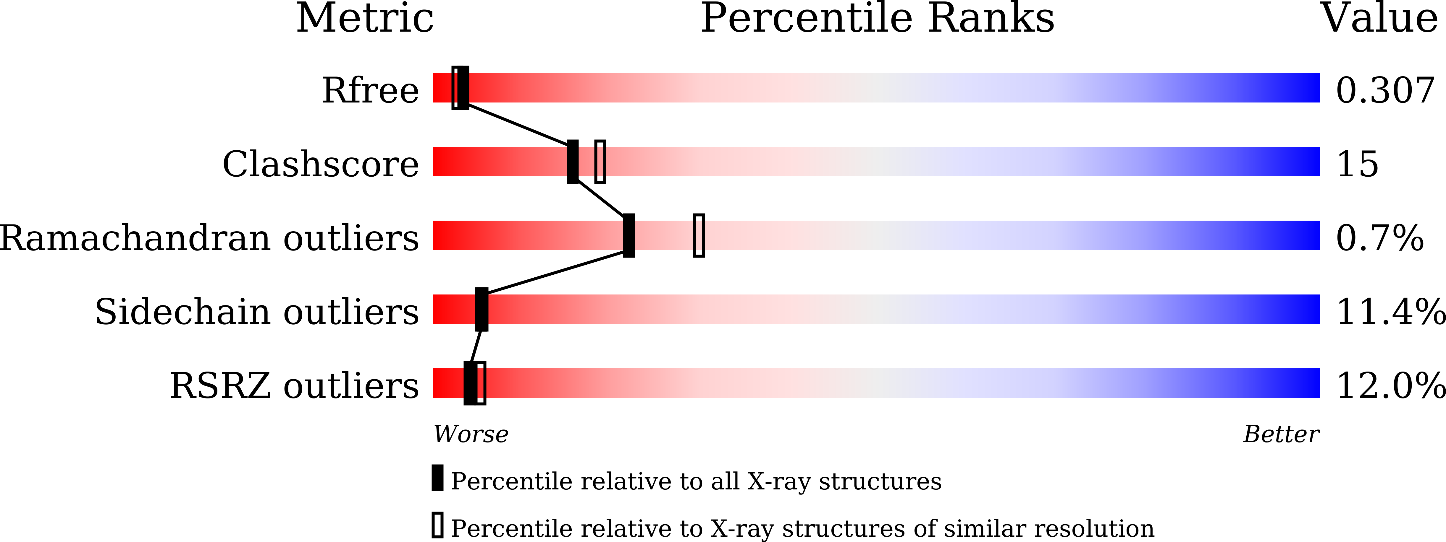
Experimental Data Snapshot
wwPDB Validation 3D Report Full Report
Entity ID: 1 | |||||
|---|---|---|---|---|---|
| Molecule | Chains | Sequence Length | Organism | Details | Image |
| 3-dehydroquinate dehydratase | 145 | Acinetobacter baumannii ATCC 17978 | Mutation(s): 0 Gene Names: aroQ, A1S_2009 EC: 4.2.1.10 | 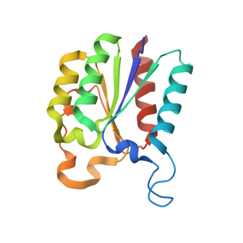 | |
UniProt | |||||
Find proteins for A3M692 (Acinetobacter baumannii (strain ATCC 17978 / CIP 53.77 / LMG 1025 / NCDC KC755 / 5377)) Explore A3M692 Go to UniProtKB: A3M692 | |||||
Entity Groups | |||||
| Sequence Clusters | 30% Identity50% Identity70% Identity90% Identity95% Identity100% Identity | ||||
| UniProt Group | A3M692 | ||||
Sequence AnnotationsExpand | |||||
| |||||
| Length ( Å ) | Angle ( ˚ ) |
|---|---|
| a = 97.993 | α = 90 |
| b = 136.254 | β = 97.59 |
| c = 143.209 | γ = 90 |
| Software Name | Purpose |
|---|---|
| REFMAC | refinement |
| HKL-2000 | data reduction |
| SCALEPACK | data scaling |
| MOLREP | phasing |
RCSB PDB (citation) is hosted by
RCSB PDB is a member of the

