5WQD

Crystal structure of TRF2 TRFH in complex with an NBS1 peptide


Experimental Data Snapshot

  • Method: X-RAY DIFFRACTION
  • Resolution: 3.00 Å
  • R-Value Free: 0.284 
  • R-Value Work: 0.233 
  • R-Value Observed: 0.235 

wwPDB Validation   3D Report Full Report


This is version 1.2 of the entry. See complete history


Literature

NBS1 Phosphorylation Status Dictates Repair Choice of Dysfunctional Telomeres

Rai, R.Hu, C.Broton, C.Chen, Y.Lei, M.Chang, S.

(2017) Mol Cell 65: 801-817.e4

  • DOI: https://doi.org/10.1016/j.molcel.2017.01.016
  • Primary Citation of Related Structures:  
    5WQD

  • PubMed Abstract: 

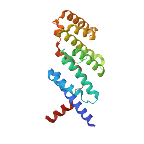
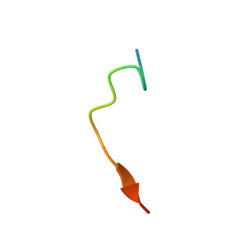
    Telomeres employ TRF2 to protect chromosome ends from activating the DNA damage sensor MRE11-RAD50-NBS1 (MRN), thereby repressing ATM-dependent DNA damage checkpoint responses. How TRF2 prevents MRN activation at dysfunctional telomeres is unclear. Here, we show that the phosphorylation status of NBS1 determines the repair pathway choice of dysfunctional telomeres. The crystal structure of the TRF2-NBS1 complex at 3.0 Å resolution shows that the NBS1 429 YQLSP 433 motif interacts specifically with the TRF2 TRFH domain. Phosphorylation of NBS1 serine 432 by CDK2 in S/G2 dissociates NBS1 from TRF2, promoting TRF2-Apollo/SNM1B complex formation and the protection of leading-strand telomeres. Classical-NHEJ-mediated repair of telomeres lacking TRF2 requires phosphorylated NBS1 S432 to activate ATM, while interaction of de-phosphorylated NBS1 S432 with TRF2 promotes alternative-NHEJ repair of telomeres lacking POT1-TPP1. Our work advances understanding of how the TRF2 TRFH domain orchestrates telomere end protection and reveals how the phosphorylation status of the NBS1 S432 dictates repair pathway choice of dysfunctional telomeres.


  • Organizational Affiliation

    Department of Laboratory Medicine, Yale University School of Medicine, 330 Cedar Street, New Haven, CT 06520, USA.


Macromolecules
Find similar proteins by:  (by identity cutoff)  |  3D Structure
Entity ID: 1
MoleculeChains Sequence LengthOrganismDetailsImage
Telomeric repeat-binding factor 2
A, B, C, D, E
A, B, C, D, E, F, G
204Homo sapiensMutation(s): 0 
Gene Names: TERF2TRBF2TRF2
UniProt & NIH Common Fund Data Resources
Find proteins for Q15554 (Homo sapiens)
Explore Q15554 
Go to UniProtKB:  Q15554
PHAROS:  Q15554
GTEx:  ENSG00000132604 
Entity Groups  
Sequence Clusters30% Identity50% Identity70% Identity90% Identity95% Identity100% Identity
UniProt GroupQ15554
Sequence Annotations
Expand
  • Reference Sequence

Find similar proteins by:  Sequence   |   3D Structure  

Entity ID: 2
MoleculeChains Sequence LengthOrganismDetailsImage
Nibrin
H, I, J, K, L
H, I, J, K, L, M, N
16Homo sapiensMutation(s): 0 
UniProt & NIH Common Fund Data Resources
Find proteins for O60934 (Homo sapiens)
Explore O60934 
Go to UniProtKB:  O60934
PHAROS:  O60934
GTEx:  ENSG00000104320 
Entity Groups  
Sequence Clusters30% Identity50% Identity70% Identity90% Identity95% Identity100% Identity
UniProt GroupO60934
Sequence Annotations
Expand
  • Reference Sequence
Experimental Data & Validation

Experimental Data

  • Method: X-RAY DIFFRACTION
  • Resolution: 3.00 Å
  • R-Value Free: 0.284 
  • R-Value Work: 0.233 
  • R-Value Observed: 0.235 
  • Space Group: P 21 21 2
Unit Cell:
Length ( Å )Angle ( ˚ )
a = 144.616α = 90
b = 153.267β = 90
c = 108.029γ = 90
Software Package:
Software NamePurpose
PHENIXrefinement
HKL-2000data collection
HKL-2000data processing
PHASERphasing
PDB_EXTRACTdata extraction
HKL-2000data reduction
HKL-2000data scaling
HKLdata reduction
HKLdata scaling

Structure Validation

View Full Validation Report


Entry History 

Deposition Data

Revision History  (Full details and data files)

  • Version 1.0: 2017-03-08
    Type: Initial release
  • Version 1.1: 2017-03-15
    Changes: Database references
  • Version 1.2: 2023-11-08
    Changes: Data collection, Database references, Refinement description