5WHF

Crystal structure of vimentin coil 1B packed in a high-order filamentous form


Experimental Data Snapshot

  • Method: X-RAY DIFFRACTION
  • Resolution: 2.25 Å
  • R-Value Free: 0.282 
  • R-Value Work: 0.235 
  • R-Value Observed: 0.237 

wwPDB Validation   3D Report Full Report


This is version 1.4 of the entry. See complete history


Literature

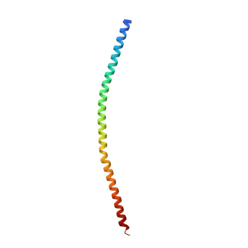
A crystal structure of coil 1B of vimentin in the filamentous form provides a model of a high-order assembly of a vimentin filament.

Pang, A.H.Obiero, J.M.Kulczyk, A.W.Sviripa, V.M.Tsodikov, O.V.

(2018) FEBS J 285: 2888-2899

  • DOI: https://doi.org/10.1111/febs.14585
  • Primary Citation of Related Structures:  
    5WHF

  • PubMed Abstract: 

    Vimentin is an intermediate filament (IF) protein that is expressed in leukocytes, fibroblasts and endothelial cells of blood vessels. Vimentin filaments contribute to structural stability of the cell membrane, organelle positioning and protein transport. Vimentin self-assembles into a dimer that subsequently forms high-order structures, including tetramers and octamers. The details of IF assembly at crystallographic resolutions are limited to the tetrameric form. We describe a crystal structure of a fragment of a vimentin rod domain (coil 1B) with a dimer of tetramers in the asymmetric unit. Coil 1B in the crystal is in an infinitely high-order filamentous assembly state, in which the tetramers are packed against each other laterally in an antiparallel fashion across the crystal lattice. In one of the directions of lateral packing, the tetramers pack against each other strictly head-to-tail, and in the orthogonal direction the tetramers pack in a staggered manner. This organization of the tetramers of coil 1B in the crystal lattice, together with previously reported biochemical and structural data, yield a model of high-order vimentin filament assembly.


  • Organizational Affiliation

    Department of Pharmaceutical Sciences, College of Pharmacy, University of Kentucky, Lexington, KY, USA.


Macromolecules
Find similar proteins by:  (by identity cutoff)  |  3D Structure
Entity ID: 1
MoleculeChains Sequence LengthOrganismDetailsImage
Vimentin
A, B, C, D, E
A, B, C, D, E, F, G, H
90Homo sapiensMutation(s): 0 
Gene Names: VIM
UniProt & NIH Common Fund Data Resources
Find proteins for P08670 (Homo sapiens)
Explore P08670 
Go to UniProtKB:  P08670
PHAROS:  P08670
GTEx:  ENSG00000026025 
Entity Groups  
Sequence Clusters30% Identity50% Identity70% Identity90% Identity95% Identity100% Identity
UniProt GroupP08670
Sequence Annotations
Expand
  • Reference Sequence
Small Molecules
Ligands 1 Unique
IDChains Name / Formula / InChI Key2D Diagram3D Interactions
GOL
Query on GOL

Download Ideal Coordinates CCD File 
I [auth G]GLYCEROL
C3 H8 O3
PEDCQBHIVMGVHV-UHFFFAOYSA-N
Experimental Data & Validation

Experimental Data

  • Method: X-RAY DIFFRACTION
  • Resolution: 2.25 Å
  • R-Value Free: 0.282 
  • R-Value Work: 0.235 
  • R-Value Observed: 0.237 
  • Space Group: P 1 21 1
Unit Cell:
Length ( Å )Angle ( ˚ )
a = 39.99α = 90
b = 77.27β = 95.03
c = 123.39γ = 90
Software Package:
Software NamePurpose
PHENIXrefinement
MOSFLMdata reduction
Aimlessdata scaling
MOLREPphasing

Structure Validation

View Full Validation Report


Entry History & Funding Information

Deposition Data


Funding OrganizationLocationGrant Number
National Institutes of Health/National Cancer Institute (NIH/NCI)United StatesCA60872

Revision History  (Full details and data files)

  • Version 1.0: 2018-06-20
    Type: Initial release
  • Version 1.1: 2018-06-27
    Changes: Data collection, Database references
  • Version 1.2: 2018-08-29
    Changes: Data collection, Database references
  • Version 1.3: 2019-12-04
    Changes: Author supporting evidence
  • Version 1.4: 2023-10-04
    Changes: Data collection, Database references, Refinement description