The entropic force generated by intrinsically disordered segments tunes protein function.
Keul, N.D., Oruganty, K., Schaper Bergman, E.T., Beattie, N.R., McDonald, W.E., Kadirvelraj, R., Gross, M.L., Phillips, R.S., Harvey, S.C., Wood, Z.A.(2018) Nature 563: 584-588
- PubMed: 30420606
- DOI: https://doi.org/10.1038/s41586-018-0699-5
- Primary Citation of Related Structures:
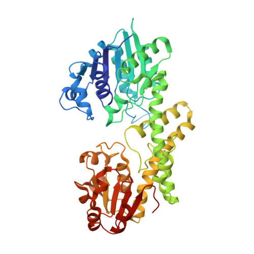
5VR8, 5W4X - PubMed Abstract:
Protein structures are dynamic and can explore a large conformational landscape 1,2 . Only some of these structural substates are important for protein function (such as ligand binding, catalysis and regulation) 3-5 . How evolution shapes the structural ensemble to optimize a specific function is poorly understood 3,4 . One of the constraints on the evolution of proteins is the stability of the folded 'native' state. Despite this, 44% of the human proteome contains intrinsically disordered peptide segments greater than 30 residues in length 6 , the majority of which have no known function 7-9 . Here we show that the entropic force produced by an intrinsically disordered carboxy terminus (ID-tail) shifts the conformational ensemble of human UDP-α-D-glucose-6-dehydrogenase (UGDH) towards a substate with a high affinity for an allosteric inhibitor. The function of the ID-tail does not depend on its sequence or chemical composition. Instead, the affinity enhancement can be accurately predicted based on the length of the intrinsically disordered segment, and is consistent with the entropic force generated by an unstructured peptide attached to the protein surface 10-13 . Our data show that the unfolded state of the ID-tail rectifies the dynamics and structure of UGDH to favour inhibitor binding. Because this entropic rectifier does not have any sequence or structural constraints, it is an easily acquired adaptation. This model implies that evolution selects for disordered segments to tune the energy landscape of proteins, which may explain the persistence of intrinsic disorder in the proteome.
Organizational Affiliation:
Department of Biochemistry and Molecular Biology, University of Georgia, Athens, GA, USA.