5VMD

Crystal structure of UBR-box from UBR6 in a domain-swapping conformation


Experimental Data Snapshot

  • Method: X-RAY DIFFRACTION
  • Resolution: 2.20 Å
  • R-Value Free: 0.247 
  • R-Value Work: 0.195 
  • R-Value Observed: 0.197 

wwPDB Validation   3D Report Full Report


This is version 1.5 of the entry. See complete history


Literature

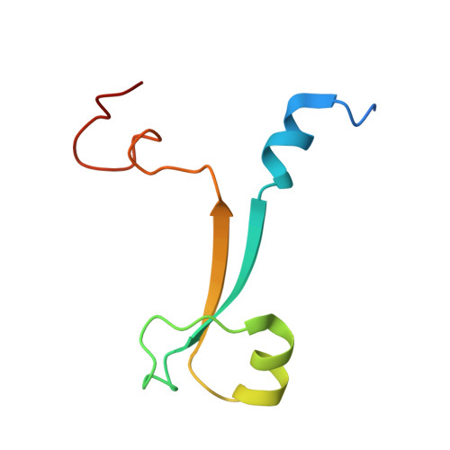
Crystal structure of the UBR-box from UBR6/FBXO11 reveals domain swapping mediated by zinc binding.

Munoz-Escobar, J.Kozlov, G.Gehring, K.

(2017) Protein Sci 26: 2092-2097

  • DOI: https://doi.org/10.1002/pro.3227
  • Primary Citation of Related Structures:  
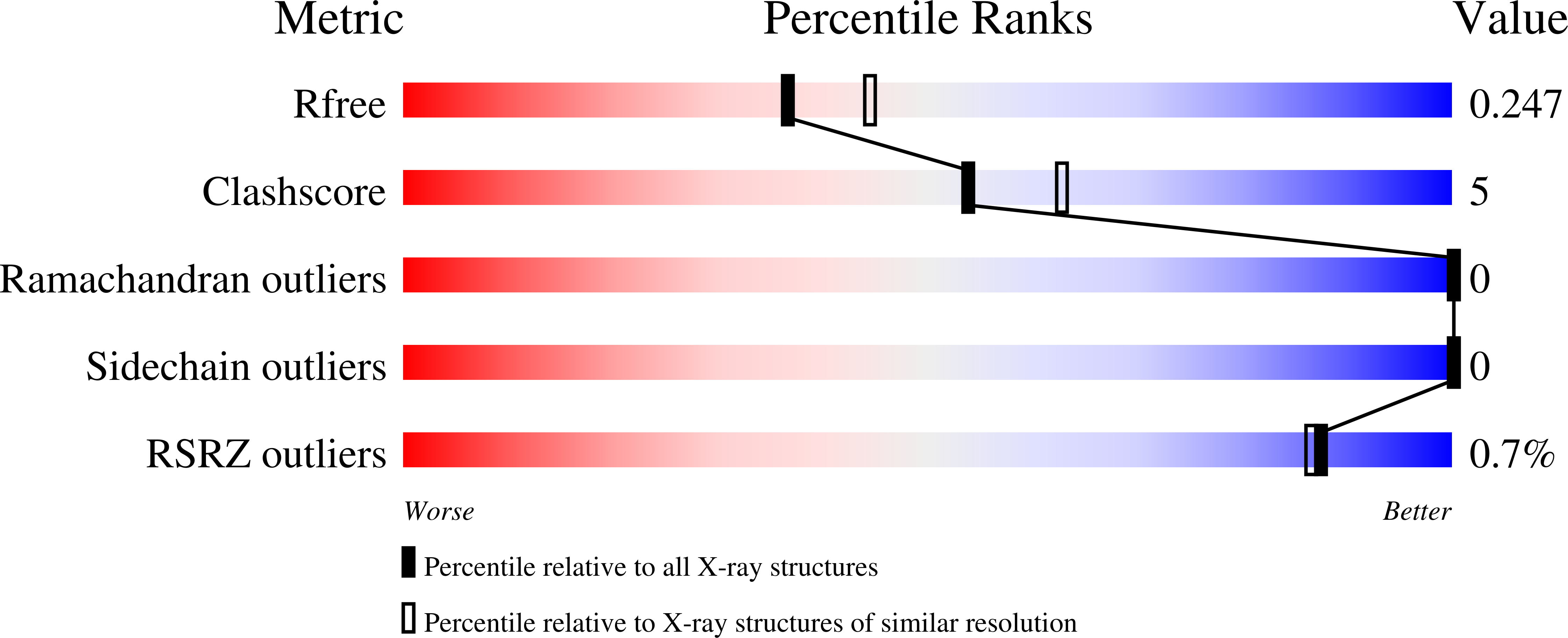
    5VMD

  • PubMed Abstract: 

    The UBR-box is a 70-residue zinc finger domain present in the UBR family of E3 ubiquitin ligases that directly binds N-terminal degradation signals in substrate proteins. UBR6, also called FBXO11, is an UBR-box containing E3 ubiquitin ligase that does not bind N-terminal signals. Here, we present the crystal structure of the UBR-box domain from human UBR6. The dimeric crystal structure reveals a unique form of domain swapping mediated by zinc coordination, where three independent protein chains come together to regenerate the topology of the monomeric UBR-box fold. Analysis of the structure suggests that the absence of N-terminal residue binding arises from the lack of an amino acid binding pocket.


  • Organizational Affiliation

    Department of Biochemistry, Groupe de Recherche Axé sur la Structure des Protéines, McGill University, Montreal, Quebec, H3G0B1, Canada.


Macromolecules
Find similar proteins by:  (by identity cutoff)  |  3D Structure
Entity ID: 1
MoleculeChains Sequence LengthOrganismDetailsImage
F-box only protein 11
A, B, C, D
77Homo sapiensMutation(s): 0 
Gene Names: FBXO11FBX11PRMT9VIT1UG063H01
UniProt & NIH Common Fund Data Resources
Find proteins for Q86XK2 (Homo sapiens)
Explore Q86XK2 
Go to UniProtKB:  Q86XK2
PHAROS:  Q86XK2
GTEx:  ENSG00000138081 
Entity Groups  
Sequence Clusters30% Identity50% Identity70% Identity90% Identity95% Identity100% Identity
UniProt GroupQ86XK2
Sequence Annotations
Expand
  • Reference Sequence
Small Molecules
Ligands 2 Unique
IDChains Name / Formula / InChI Key2D Diagram3D Interactions
ZN
Query on ZN

Download Ideal Coordinates CCD File 
E [auth A]
F [auth A]
G [auth A]
H [auth A]
I [auth A]
E [auth A],
F [auth A],
G [auth A],
H [auth A],
I [auth A],
N [auth B],
O [auth B],
P [auth B],
Q [auth B],
R [auth B],
U [auth C],
V [auth C],
X [auth D],
Y [auth D]
ZINC ION
Zn
PTFCDOFLOPIGGS-UHFFFAOYSA-N
EDO
Query on EDO

Download Ideal Coordinates CCD File 
AA [auth D]
J [auth A]
K [auth A]
L [auth A]
M [auth A]
AA [auth D],
J [auth A],
K [auth A],
L [auth A],
M [auth A],
S [auth B],
T [auth B],
W [auth C],
Z [auth D]
1,2-ETHANEDIOL
C2 H6 O2
LYCAIKOWRPUZTN-UHFFFAOYSA-N
Experimental Data & Validation

Experimental Data

  • Method: X-RAY DIFFRACTION
  • Resolution: 2.20 Å
  • R-Value Free: 0.247 
  • R-Value Work: 0.195 
  • R-Value Observed: 0.197 
  • Space Group: P 21 21 21
Unit Cell:
Length ( Å )Angle ( ˚ )
a = 67.57α = 90
b = 69.2β = 90
c = 82.59γ = 90
Software Package:
Software NamePurpose
PHENIXrefinement
HKL-2000data reduction
HKL-2000data scaling
PHENIXphasing

Structure Validation

View Full Validation Report


Entry History & Funding Information

Deposition Data


Funding OrganizationLocationGrant Number
Canadian Institutes of Health Research (CIHR)Canada--

Revision History  (Full details and data files)

  • Version 1.0: 2017-07-12
    Type: Initial release
  • Version 1.1: 2017-07-26
    Changes: Author supporting evidence, Database references
  • Version 1.2: 2017-08-09
    Changes: Database references
  • Version 1.3: 2017-10-04
    Changes: Database references
  • Version 1.4: 2020-01-08
    Changes: Author supporting evidence
  • Version 1.5: 2024-03-13
    Changes: Data collection, Database references, Derived calculations, Refinement description