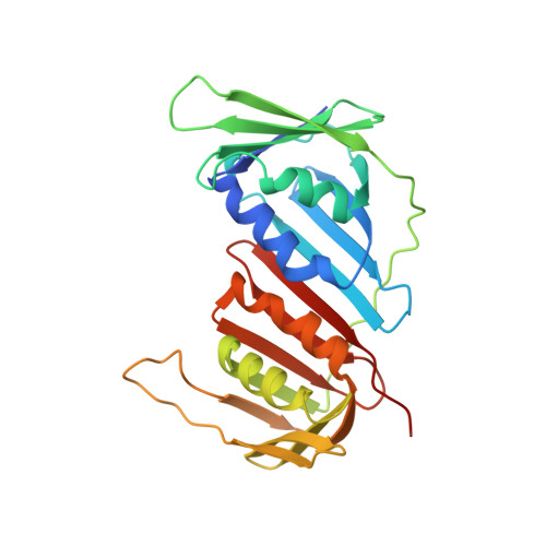
Crystal structures of PCNA mutant proteins defective in gene silencing suggest a novel interaction site on the front face of the PCNA ring.
Kondratick, C.M., Litman, J.M., Shaffer, K.V., Washington, M.T., Dieckman, L.M.(2018) PLoS One 13: e0193333-e0193333
- PubMed: 29499038
- DOI: https://doi.org/10.1371/journal.pone.0193333
- Primary Citation of Related Structures:
5V7K, 5V7L, 5V7M - PubMed Abstract:
Proliferating cell nuclear antigen (PCNA), a homotrimeric protein, is the eukaryotic sliding clamp that functions as a processivity factor for polymerases during DNA replication. Chromatin association factor 1 (CAF-1) is a heterotrimeric histone chaperone protein that is required for coupling chromatin assembly with DNA replication in eukaryotes. CAF-1 association with replicating DNA, and the targeting of newly synthesized histones to sites of DNA replication and repair requires its interaction with PCNA. Genetic studies have identified three mutant forms of PCNA in yeast that cause defects in gene silencing and exhibit altered association of CAF-1 to chromatin in vivo, as well as inhibit binding to CAF-1 in vitro. Three of these mutant forms of PCNA, encoded by the pol30-6, pol30-8, and the pol30-79 alleles, direct the synthesis of PCNA proteins with the amino acid substitutions D41A/D42A, R61A/D63A, and L126A/I128A, respectively. Interestingly, these double alanine substitutions are located far away from each other within the PCNA protein. To understand the structural basis of the interaction between PCNA and CAF-1 and how disruption of this interaction leads to reduced gene silencing, we determined the X-ray crystal structures of each of these mutant PCNA proteins. All three of the substitutions caused disruptions of a surface cavity on the front face of the PCNA ring, which is formed in part by three loops comprised of residues 21-24, 41-44, and 118-134. We suggest that this cavity is a novel binding pocket required for the interaction between PCNA and CAF-1, and that this region in PCNA also represents a potential binding site for other PCNA-binding proteins.
Organizational Affiliation:
Department of Biochemistry, University of Iowa College of Medicine, Iowa City, IA, United States of America.