5UKW

Crystal structure of human Glucose 6-phosphate Dehydrogenase mutant (A277C) complexed with G6P


Experimental Data Snapshot

  • Method: X-RAY DIFFRACTION
  • Resolution: 2.65 Å
  • R-Value Free: 0.234 
  • R-Value Work: 0.189 
  • R-Value Observed: 0.191 

wwPDB Validation   3D Report Full Report


Ligand Structure Quality Assessment 


This is version 1.5 of the entry. See complete history


Literature

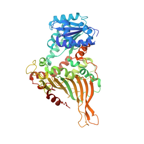
Mutations in the tetramer interface of human glucose-6-phosphate dehydrogenase reveals kinetic differences between oligomeric states.

Ranzani, A.T.Cordeiro, A.T.

(2017) FEBS Lett 591: 1278-1284

  • DOI: https://doi.org/10.1002/1873-3468.12638
  • Primary Citation of Related Structures:  
    5UKW

  • PubMed Abstract: 

    Glucose-6-phosphate dehydrogenase (G6PDH) catalyzes the oxidation of glucose-6-phoshate to 6-phospho-gluconolactone with the concomitant reduction of NADP + to NADPH. In solution, the recombinant human G6PDH is known to be active as dimers and tetramers. To distinguish between the kinetic properties of dimers and tetramers of the G6PDH is not trivial. Steady-state kinetic experiments are often performed at low enzyme concentrations, which favor the dimeric state. The present work describes two novel human G6PDH mutants, one that creates four disulfide bonds among apposing dimers, resulting in a 'cross-linked' tetramer, and another that prevents the dimer to dimer association. The functional and structural characterizations of such mutants indicate the tetramer as the most active form of human G6PDH.


  • Organizational Affiliation

    Institute of Biology, University of Campinas, Sao Paulo, Brazil.


Macromolecules
Find similar proteins by:  (by identity cutoff)  |  3D Structure
Entity ID: 1
MoleculeChains Sequence LengthOrganismDetailsImage
Glucose-6-phosphate 1-dehydrogenase488Homo sapiensMutation(s): 1 
Gene Names: G6PD
EC: 1.1.1.49
UniProt & NIH Common Fund Data Resources
Find proteins for P11413 (Homo sapiens)
Explore P11413 
Go to UniProtKB:  P11413
PHAROS:  P11413
GTEx:  ENSG00000160211 
Entity Groups  
Sequence Clusters30% Identity50% Identity70% Identity90% Identity95% Identity100% Identity
UniProt GroupP11413
Sequence Annotations
Expand
  • Reference Sequence
Small Molecules
Ligands 2 Unique
IDChains Name / Formula / InChI Key2D Diagram3D Interactions
BG6
Query on BG6

Download Ideal Coordinates CCD File 
B [auth A]6-O-phosphono-beta-D-glucopyranose
C6 H13 O9 P
NBSCHQHZLSJFNQ-VFUOTHLCSA-N
GOL
Query on GOL

Download Ideal Coordinates CCD File 
C [auth A]GLYCEROL
C3 H8 O3
PEDCQBHIVMGVHV-UHFFFAOYSA-N
Experimental Data & Validation

Experimental Data

  • Method: X-RAY DIFFRACTION
  • Resolution: 2.65 Å
  • R-Value Free: 0.234 
  • R-Value Work: 0.189 
  • R-Value Observed: 0.191 
  • Space Group: F 2 2 2
Unit Cell:
Length ( Å )Angle ( ˚ )
a = 59.099α = 90
b = 177.817β = 90
c = 216.47γ = 90
Software Package:
Software NamePurpose
REFMACrefinement
XDSdata reduction
Aimlessdata scaling
MOLREPphasing

Structure Validation

View Full Validation Report


Ligand Structure Quality Assessment 


Entry History & Funding Information

Deposition Data


Funding OrganizationLocationGrant Number
Sao Paulo Research Foundation (FAPESP)Brazil2012/23682-7

Revision History  (Full details and data files)

  • Version 1.0: 2017-04-19
    Type: Initial release
  • Version 1.1: 2017-05-10
    Changes: Database references
  • Version 1.2: 2019-04-17
    Changes: Author supporting evidence, Data collection
  • Version 1.3: 2020-01-01
    Changes: Author supporting evidence
  • Version 1.4: 2020-07-29
    Type: Remediation
    Reason: Carbohydrate remediation
    Changes: Data collection, Derived calculations, Structure summary
  • Version 1.5: 2023-10-04
    Changes: Data collection, Database references, Refinement description, Structure summary