5TXW

Structure of Pfp1 protease from Thermococcus thioreducens: large cell H3 crystal form


Experimental Data Snapshot

  • Method: X-RAY DIFFRACTION
  • Resolution: 1.86 Å
  • R-Value Free: 0.173 
  • R-Value Work: 0.127 
  • R-Value Observed: 0.130 

wwPDB Validation   3D Report Full Report


This is version 1.4 of the entry. See complete history


Literature

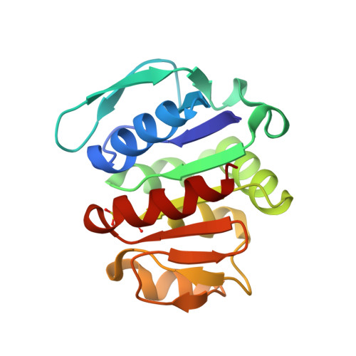
The structure of the Pfp1 protease from the hyperthermophilic archaeon Thermococcus thioreducens in two crystal forms.

Larson, S.B.McPherson, A.

(2017) Acta Crystallogr D Struct Biol 73: 749-756

  • DOI: https://doi.org/10.1107/S2059798317010622
  • Primary Citation of Related Structures:  
    5TW0, 5TXW

  • PubMed Abstract: 

    The Pfp1 protease, a cysteine protease of unknown specificity from the hyperthermophilic archaeon Thermococcus thioreducens, was crystallized in two distinctive crystal forms: from concentrated citrate in one case and PEG in the other. X-ray data were collected from both crystal forms at room temperature to about 1.9 Å resolution using a laboratory source and detector, and the structures were solved by molecular replacement using the Pfp1 protease from Pyrococcus horikoshii as the search model. In the T. thioreducens protease structures, Cys18 residues on adjacent molecules in the asymmetric units form intermolecular disulfide bonds, thereby yielding hexamers composed of three cross-linked, quasi-dyad-related dimers with crystallographically exact threefold axes and exhibiting almost exact 32 symmetry. The corresponding residue in P. horikoshii Pfp1 is Tyr18. An individual active site containing Cys100 and His101 also includes a Glu74 residue contributed by a quasi-twofold-related, non-cross-linked subunit. Two catalytic triads are therefore closely juxtaposed about the quasi-twofold axis at the interface of these subunits, and are relatively sequestered within the hexamer cavity. The cysteine in the active site is observed to be oxidized in both of the crystal forms that were studied.


  • Organizational Affiliation

    Department of Molecular Biology and Biochemistry, University of California, 530A Steinhaus Hall, Irvine, CA 92697-3900, USA.


Macromolecules
Find similar proteins by:  (by identity cutoff)  |  3D Structure
Entity ID: 1
MoleculeChains Sequence LengthOrganismDetailsImage
Peptidase
A, B, C, D
166Thermococcus thioreducensMutation(s): 0 
Gene Names: AMR53_10535
UniProt
Find proteins for A0A0Q2XKL6 (Thermococcus thioreducens)
Explore A0A0Q2XKL6 
Go to UniProtKB:  A0A0Q2XKL6
Entity Groups  
Sequence Clusters30% Identity50% Identity70% Identity90% Identity95% Identity100% Identity
UniProt GroupA0A0Q2XKL6
Sequence Annotations
Expand
  • Reference Sequence
Small Molecules
Modified Residues  1 Unique
IDChains TypeFormula2D DiagramParent
OCS
Query on OCS
A, B, C, D
L-PEPTIDE LINKINGC3 H7 N O5 SCYS
Experimental Data & Validation

Experimental Data

  • Method: X-RAY DIFFRACTION
  • Resolution: 1.86 Å
  • R-Value Free: 0.173 
  • R-Value Work: 0.127 
  • R-Value Observed: 0.130 
  • Space Group: H 3
Unit Cell:
Length ( Å )Angle ( ˚ )
a = 152.85α = 90
b = 152.85β = 90
c = 82.541γ = 120
Software Package:
Software NamePurpose
iMOSFLMdata reduction
Aimlessdata scaling
CNSphasing
REFMACrefinement
Cootmodel building

Structure Validation

View Full Validation Report


Entry History 

Deposition Data

Revision History  (Full details and data files)

  • Version 1.0: 2017-09-13
    Type: Initial release
  • Version 1.1: 2017-11-22
    Changes: Database references
  • Version 1.2: 2018-04-18
    Changes: Data collection, Database references
  • Version 1.3: 2023-10-04
    Changes: Data collection, Database references, Refinement description
  • Version 1.4: 2023-11-15
    Changes: Data collection