5TCX

Crystal structure of human tetraspanin CD81


Experimental Data Snapshot

  • Method: X-RAY DIFFRACTION
  • Resolution: 2.96 Å
  • R-Value Free: 0.293 
  • R-Value Work: 0.267 
  • R-Value Observed: 0.270 

wwPDB Validation   3D Report Full Report


Ligand Structure Quality Assessment 


This is version 1.4 of the entry. See complete history


Literature

Crystal Structure of a Full-Length Human Tetraspanin Reveals a Cholesterol-Binding Pocket.

Zimmerman, B.Kelly, B.McMillan, B.J.Seegar, T.C.Dror, R.O.Kruse, A.C.Blacklow, S.C.

(2016) Cell 167: 1041-1051.e11

  • DOI: https://doi.org/10.1016/j.cell.2016.09.056
  • Primary Citation of Related Structures:  
    5TCX

  • PubMed Abstract: 

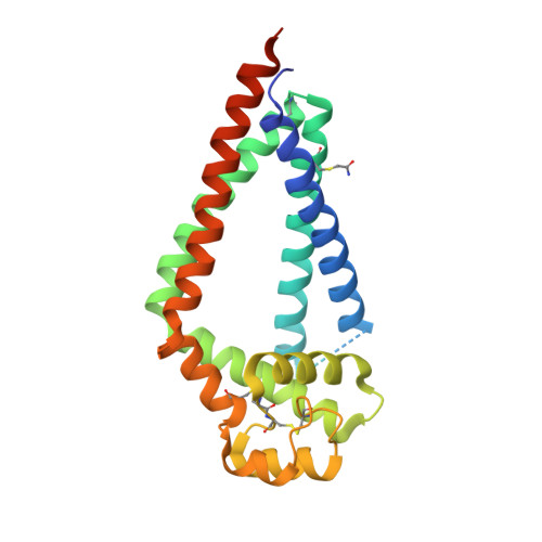
    Tetraspanins comprise a diverse family of four-pass transmembrane proteins that play critical roles in the immune, reproductive, genitourinary, and auditory systems. Despite their pervasive roles in human physiology, little is known about the structure of tetraspanins or the molecular mechanisms underlying their various functions. Here, we report the crystal structure of human CD81, a full-length tetraspanin. The transmembrane segments of CD81 pack as two largely separated pairs of helices, capped by the large extracellular loop (EC2) at the outer membrane leaflet. The two pairs of helices converge at the inner leaflet to create an intramembrane pocket with additional electron density corresponding to a bound cholesterol molecule within the cavity. Molecular dynamics simulations identify an additional conformation in which EC2 separates substantially from the transmembrane domain. Cholesterol binding appears to modulate CD81 activity in cells, suggesting a potential mechanism for regulation of tetraspanin function.


  • Organizational Affiliation

    Department of Biological Chemistry and Molecular Pharmacology, Harvard Medical School, Boston, MA 02115, USA; Department of Cancer Biology, Dana Farber Cancer Institute, Boston, MA 02215, USA.


Macromolecules
Find similar proteins by:  (by identity cutoff)  |  3D Structure
Entity ID: 1
MoleculeChains Sequence LengthOrganismDetailsImage
CD81 antigen246Homo sapiensMutation(s): 4 
Gene Names: CD81TAPA1TSPAN28
Membrane Entity: Yes 
UniProt & NIH Common Fund Data Resources
Find proteins for P60033 (Homo sapiens)
Explore P60033 
Go to UniProtKB:  P60033
PHAROS:  P60033
GTEx:  ENSG00000110651 
Entity Groups  
Sequence Clusters30% Identity50% Identity70% Identity90% Identity95% Identity100% Identity
UniProt GroupP60033
Sequence Annotations
Expand
  • Reference Sequence
Small Molecules
Ligands 1 Unique
IDChains Name / Formula / InChI Key2D Diagram3D Interactions
CLR
Query on CLR

Download Ideal Coordinates CCD File 
B [auth A]CHOLESTEROL
C27 H46 O
HVYWMOMLDIMFJA-DPAQBDIFSA-N
Modified Residues  1 Unique
IDChains TypeFormula2D DiagramParent
YCM
Query on YCM
A
L-PEPTIDE LINKINGC5 H10 N2 O3 SCYS
Experimental Data & Validation

Experimental Data

  • Method: X-RAY DIFFRACTION
  • Resolution: 2.96 Å
  • R-Value Free: 0.293 
  • R-Value Work: 0.267 
  • R-Value Observed: 0.270 
  • Space Group: C 1 2 1
Unit Cell:
Length ( Å )Angle ( ˚ )
a = 101.43α = 90
b = 73.51β = 125.11
c = 70.35γ = 90
Software Package:
Software NamePurpose
PHENIXrefinement
XDSdata reduction
XDSdata scaling
PHASERphasing
Cootmodel building

Structure Validation

View Full Validation Report


Ligand Structure Quality Assessment 


Entry History & Funding Information

Deposition Data


Funding OrganizationLocationGrant Number
National Institutes of Health/National Cancer Institute (NIH/NCI)United States5RO1 CA092433
National Institutes of Health/Office of the DirectorUnited States1DP5 OD021345

Revision History  (Full details and data files)

  • Version 1.0: 2016-11-09
    Type: Initial release
  • Version 1.1: 2016-12-21
    Changes: Database references
  • Version 1.2: 2017-09-27
    Changes: Author supporting evidence
  • Version 1.3: 2019-12-04
    Changes: Author supporting evidence
  • Version 1.4: 2023-10-04
    Changes: Data collection, Database references, Refinement description