5T30

Human MnSOD-azide complex


Experimental Data Snapshot

  • Method: X-RAY DIFFRACTION
  • Resolution: 1.77 Å
  • R-Value Free: 0.239 
  • R-Value Work: 0.209 
  • R-Value Observed: 0.210 

wwPDB Validation   3D Report Full Report


This is version 1.3 of the entry. See complete history


Literature

Substrate-analog binding and electrostatic surfaces of human manganese superoxide dismutase.

Azadmanesh, J.Trickel, S.R.Borgstahl, G.E.O.

(2017) J Struct Biol 199: 68-75

  • DOI: https://doi.org/10.1016/j.jsb.2017.04.011
  • Primary Citation of Related Structures:  
    5T30, 5VF9

  • PubMed Abstract: 

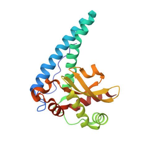
    Superoxide dismutases (SODs) are enzymes that play a key role in protecting cells from toxic oxygen metabolites by disproportionation of two molecules of superoxide into molecular oxygen and hydrogen peroxide via cyclic reduction and oxidation at the active site metal. The azide anion is a potent competitive inhibitor that binds directly to the metal and is used as a substrate analog to superoxide in studies of SOD. The crystal structure of human MnSOD-azide complex was solved and shows the putative binding position of superoxide, providing a model for binding to the active site. Azide is bound end-on at the sixth coordinate position of the manganese ion. Tetrameric electrostatic surfaces were calculated incorporating accurate partial charges for the active site in three states, including a state with superoxide coordinated to the metal using the position of azide as a model. These show facilitation of the anionic ligand to the active site pit via a 'valley' of positively-charged surface patches. Surrounding ridges of negative charge help guide the superoxide anion. Within the active site pit, Arg173 and Glu162 further guide and align superoxide for efficient catalysis. Superoxide coordination at the sixth position causes the electrostatic surface of the active site pit to become nearly neutral. A model for electrostatic-mediated diffusion, and efficient binding of superoxide for catalysis is presented.


  • Organizational Affiliation

    The Eppley Institute for Research in Cancer and Allied Diseases, 987696 Nebraska Medical Center, Omaha, NE 68198-7696, USA; Department of Biochemistry and Molecular Biology, 985870 Nebraska Medical Center, Omaha, NE 68198-5870, USA.


Macromolecules
Find similar proteins by:  (by identity cutoff)  |  3D Structure
Entity ID: 1
MoleculeChains Sequence LengthOrganismDetailsImage
Superoxide dismutase [Mn], mitochondrial
A, B
199Homo sapiensMutation(s): 0 
Gene Names: SOD2
EC: 1.15.1.1
UniProt & NIH Common Fund Data Resources
Find proteins for P04179 (Homo sapiens)
Explore P04179 
Go to UniProtKB:  P04179
PHAROS:  P04179
Entity Groups  
Sequence Clusters30% Identity50% Identity70% Identity90% Identity95% Identity100% Identity
UniProt GroupP04179
Sequence Annotations
Expand
  • Reference Sequence
Small Molecules
Ligands 4 Unique
IDChains Name / Formula / InChI Key2D Diagram3D Interactions
PO4
Query on PO4

Download Ideal Coordinates CCD File 
D [auth A],
I [auth B]
PHOSPHATE ION
O4 P
NBIIXXVUZAFLBC-UHFFFAOYSA-K
MN
Query on MN

Download Ideal Coordinates CCD File 
C [auth A],
H [auth B]
MANGANESE (II) ION
Mn
WAEMQWOKJMHJLA-UHFFFAOYSA-N
AZI
Query on AZI

Download Ideal Coordinates CCD File 
G [auth A]AZIDE ION
N3
IVRMZWNICZWHMI-UHFFFAOYSA-N
K
Query on K

Download Ideal Coordinates CCD File 
E [auth A],
F [auth A]
POTASSIUM ION
K
NPYPAHLBTDXSSS-UHFFFAOYSA-N
Experimental Data & Validation

Experimental Data

  • Method: X-RAY DIFFRACTION
  • Resolution: 1.77 Å
  • R-Value Free: 0.239 
  • R-Value Work: 0.209 
  • R-Value Observed: 0.210 
  • Space Group: P 61 2 2
Unit Cell:
Length ( Å )Angle ( ˚ )
a = 77.9α = 90
b = 77.9β = 90
c = 238.442γ = 120
Software Package:
Software NamePurpose
REFMACrefinement
Cootmodel building

Structure Validation

View Full Validation Report


Entry History & Funding Information

Deposition Data


Funding OrganizationLocationGrant Number
National Aeronautic Space Administration (NASA, United States)United StatesNNX15AM82A

Revision History  (Full details and data files)

  • Version 1.0: 2017-05-17
    Type: Initial release
  • Version 1.1: 2017-06-28
    Changes: Database references
  • Version 1.2: 2020-01-01
    Changes: Author supporting evidence
  • Version 1.3: 2024-03-06
    Changes: Data collection, Database references, Derived calculations, Refinement description