The Role of Disulfide Bond Replacements in Analogues of the Tarantula Toxin ProTx-II and Their Effects on Inhibition of the Voltage-Gated Sodium Ion Channel Nav1.7.
Wright, Z.V.F., McCarthy, S., Dickman, R., Reyes, F.E., Sanchez-Martinez, S., Cryar, A., Kilford, I., Hall, A., Takle, A.K., Topf, M., Gonen, T., Thalassinos, K., Tabor, A.B.(2017) J Am Chem Soc 139: 13063-13075
- PubMed: 28880078
- DOI: https://doi.org/10.1021/jacs.7b06506
- Primary Citation of Related Structures:
5O0U - PubMed Abstract:
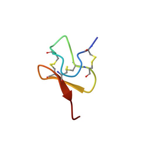
Spider venom toxins, such as Protoxin-II (ProTx-II), have recently received much attention as selective Na v 1.7 channel blockers, with potential to be developed as leads for the treatment of chronic nocioceptive pain. ProTx-II is a 30-amino acid peptide with three disulfide bonds that has been reported to adopt a well-defined inhibitory cystine knot (ICK) scaffold structure. Potential drawbacks with such peptides include poor pharmacodynamics and potential scrambling of the disulfide bonds in vivo. In order to address these issues, in the present study we report the solid-phase synthesis of lanthionine-bridged analogues of ProTx-II, in which one of the three disulfide bridges is replaced with a thioether linkage, and evaluate the biological properties of these analogues. We have also investigated the folding and disulfide bridging patterns arising from different methods of oxidation of the linear peptide precursor. Finally, we report the X-ray crystal structure of ProTx-II to atomic resolution; to our knowledge this is the first crystal structure of an ICK spider venom peptide not bound to a substrate.
Organizational Affiliation:
Department of Chemistry, University College London , 20 Gordon Street, London WC1H 0AJ, United Kingdom.


