Structure and oligomerization of the periplasmic domain of GspL from the type II secretion system of Pseudomonas aeruginosa.
Fulara, A., Vandenberghe, I., Read, R.J., Devreese, B., Savvides, S.N.(2018) Sci Rep 8: 16760-16760
- PubMed: 30425318
- DOI: https://doi.org/10.1038/s41598-018-34956-w
- Primary Citation of Related Structures:
5N7L, 6GHU - PubMed Abstract:
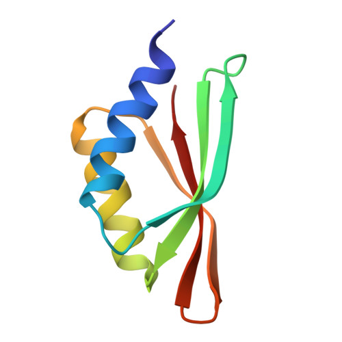
The ability of bacteria to infect a host relies in part on the secretion of molecular virulence factors across the cell envelope. Pseudomonas aeruginosa, a ubiquitous environmental bacterium causing opportunistic infections in humans, employs the type II secretion system (T2SS) to transport effector proteins across its cellular envelope as part of a diverse array of virulence strategies. General secretory pathway protein L (GspL) is an essential inner-membrane component of the T2SS apparatus, and is thought to facilitate transduction of the energy from ATP hydrolysis in the cytoplasm to the periplasmic components of the system. However, our incomplete understanding of the assembly principles of the T2SS machinery prevents the mechanistic deconvolution of T2SS-mediated protein secretion. Here we show via two crystal structures that the periplasmic ferredoxin-like domain of GspL (GspL fld ) is a dimer stabilized by hydrophobic interactions, and that this interface may allow significant interdomain plasticity. The general dimerization mode of GspL fld is shared with GspL from Vibrio parahaemolyticus suggesting a conserved oligomerization mode across the GspL family. Furthermore, we identified a tetrameric form of the complete periplasmic segment of GspL (GspL peri ) which indicates that GspL may be able to adopt multiple oligomeric states as part of its dynamic role in the T2SS apparatus.
- Unit for Structural Biology, Department of Biochemistry and Microbiology, Ghent University, 9052, Ghent (Zwijnaarde), Belgium.
Organizational Affiliation: