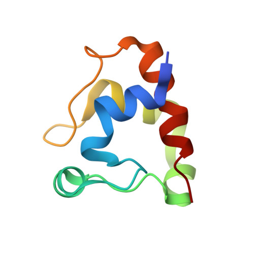
5MXY

KustC0563 c-type cytochrome


Experimental Data Snapshot

  • Method: X-RAY DIFFRACTION
  • Resolution: 1.90 Å
  • R-Value Free: 0.230 
  • R-Value Work: 0.199 
  • R-Value Observed: 0.200 

wwPDB Validation   3D Report Full Report


Ligand Structure Quality Assessment 


This is version 1.1 of the entry. See complete history


Literature

Specificity of Small c -Type Cytochromes in Anaerobic Ammonium Oxidation.

Akram, M.Bock, J.Dietl, A.Barends, T.R.M.

(2021) ACS Omega 6: 21457-21464

  • DOI: https://doi.org/10.1021/acsomega.1c02275
  • Primary Citation of Related Structures:  
    5MXY, 7O38

  • PubMed Abstract: 

    Anaerobic ammonium oxidation (anammox) is a bacterial process in which ammonium and nitrite are combined into dinitrogen gas and water, yielding energy for the cell. This process relies on a series of redox reactions catalyzed by a set of enzymes, with electrons being shuttled to and from these enzymes, likely by small cytochrome c proteins. For this system to work productively, these electron carriers require a degree of specificity toward the various possible redox partners they encounter in the cell. Here, we compare two cytochrome c proteins from the anammox model organism Kuenenia stuttgartiensis . We show that they are highly homologous, are expressed at comparable levels, share the same fold, and display highly similar redox potentials, yet one of them accepts electrons from the metabolic enzyme hydroxylamine oxidase (HAO) efficiently, whereas the other does not. An analysis of the crystal structures supplemented by Monte Carlo simulations of the transient redox interactions suggests that this difference is at least partly due to the electrostatic field surrounding the proteins, illustrating one way in which the electron carriers in anammox could attain the required specificity. Moreover, the simulations suggest a different "outlet" for electrons on HAO than has traditionally been assumed.


  • Organizational Affiliation

    Department of Biomolecular Mechanisms, Max Planck Institute for Medical Research, Jahnstrasse 29, D-69120 Heidelberg, Germany.


Macromolecules
Find similar proteins by:  (by identity cutoff)  |  3D Structure
Entity ID: 1
MoleculeChains Sequence LengthOrganismDetailsImage
Cytochrome c-552 Ks_335885Candidatus Kuenenia stuttgartiensisMutation(s): 0 
Gene Names: kustc0563
UniProt
Find proteins for Q30JB5 (Kuenenia stuttgartiensis)
Explore Q30JB5 
Go to UniProtKB:  Q30JB5
Entity Groups  
Sequence Clusters30% Identity50% Identity70% Identity90% Identity95% Identity100% Identity
UniProt GroupQ30JB5
Sequence Annotations
Expand
  • Reference Sequence
Small Molecules
Ligands 4 Unique
IDChains Name / Formula / InChI Key2D Diagram3D Interactions
HEC
Query on HEC

Download Ideal Coordinates CCD File 
B [auth A]HEME C
C34 H34 Fe N4 O4
HXQIYSLZKNYNMH-LJNAALQVSA-N
ZN
Query on ZN

Download Ideal Coordinates CCD File 
C [auth A]
D [auth A]
E [auth A]
F [auth A]
G [auth A]
C [auth A],
D [auth A],
E [auth A],
F [auth A],
G [auth A],
H [auth A],
I [auth A],
J [auth A],
K [auth A],
L [auth A]
ZINC ION
Zn
PTFCDOFLOPIGGS-UHFFFAOYSA-N
EDO
Query on EDO

Download Ideal Coordinates CCD File 
M [auth A]1,2-ETHANEDIOL
C2 H6 O2
LYCAIKOWRPUZTN-UHFFFAOYSA-N
ACT
Query on ACT

Download Ideal Coordinates CCD File 
N [auth A]ACETATE ION
C2 H3 O2
QTBSBXVTEAMEQO-UHFFFAOYSA-M
Experimental Data & Validation

Experimental Data

  • Method: X-RAY DIFFRACTION
  • Resolution: 1.90 Å
  • R-Value Free: 0.230 
  • R-Value Work: 0.199 
  • R-Value Observed: 0.200 
  • Space Group: P 43 21 2
Unit Cell:
Length ( Å )Angle ( ˚ )
a = 47.82α = 90
b = 47.82β = 90
c = 99.93γ = 90
Software Package:
Software NamePurpose
REFMACrefinement
XDSdata reduction
XSCALEdata scaling
autoSHARPphasing

Structure Validation

View Full Validation Report


Ligand Structure Quality Assessment 


Entry History 

Deposition Data

Revision History  (Full details and data files)

  • Version 1.0: 2018-02-14
    Type: Initial release
  • Version 1.1: 2021-10-13
    Changes: Data collection, Database references, Derived calculations