5MBA

BINDING MODE OF AZIDE TO FERRIC APLYSIA LIMACINA MYOGLOBIN. CRYSTALLOGRAPHIC ANALYSIS AT 1.9 ANGSTROMS RESOLUTION


Experimental Data Snapshot

  • Method: X-RAY DIFFRACTION
  • Resolution: 1.90 Å
  • R-Value Observed: 0.139 

wwPDB Validation   3D Report Full Report


Ligand Structure Quality Assessment 


This is version 1.2 of the entry. See complete history


Literature

Binding mode of azide to ferric Aplysia limacina myoglobin. Crystallographic analysis at 1.9 A resolution.

Mattevi, A.Gatti, G.Coda, A.Rizzi, M.Ascenzi, P.Brunori, M.Bolognesi, M.

(1991) J Mol Recognit 4: 1-6

  • DOI: https://doi.org/10.1002/jmr.300040102
  • Primary Citation of Related Structures:  
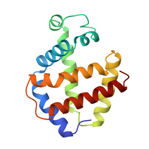
    5MBA

  • PubMed Abstract: 

    The binding mode of azide to the ferric form of Aplysia limacina myoglobin has been studied by X-ray crystallography. The three-dimensional structure of the complex has been refined at 1.9 A resolution to a crystallographic R-factor of 13.9%, including 126 ordered solvent molecules. Azide binds to the heme iron, at the sixth co-ordination position, and is oriented towards the outer part of the distal site crevice. This orientation is stabilized by an ionic interaction with the side-chain of Arg66 (E10) which, from an outer orientation in the 'aquo-met' ligand-free myoglobin, folds back towards the distal site in the presence of the anionic ligand. In the absence of a hydrogen bond donor residue at the distal E7 position in Aplysia limacina myoglobin, a different polar residue, Arg66 at the E10 topological position, has been selected by molecular evolution in order to grant ligand stabilization.


  • Organizational Affiliation

    Dipartimento di Genetica e Microbiologia, Università di Pavia, Italy.


Macromolecules
Find similar proteins by:  (by identity cutoff)  |  3D Structure
Entity ID: 1
MoleculeChains Sequence LengthOrganismDetailsImage
MYOGLOBIN147Aplysia limacinaMutation(s): 0 
UniProt
Find proteins for P02210 (Aplysia limacina)
Explore P02210 
Go to UniProtKB:  P02210
Entity Groups  
Sequence Clusters30% Identity50% Identity70% Identity90% Identity95% Identity100% Identity
UniProt GroupP02210
Sequence Annotations
Expand
  • Reference Sequence
Small Molecules
Ligands 2 Unique
IDChains Name / Formula / InChI Key2D Diagram3D Interactions
HEM
Query on HEM

Download Ideal Coordinates CCD File 
C [auth A]PROTOPORPHYRIN IX CONTAINING FE
C34 H32 Fe N4 O4
KABFMIBPWCXCRK-RGGAHWMASA-L
AZI
Query on AZI

Download Ideal Coordinates CCD File 
B [auth A]AZIDE ION
N3
IVRMZWNICZWHMI-UHFFFAOYSA-N
Experimental Data & Validation

Experimental Data

  • Method: X-RAY DIFFRACTION
  • Resolution: 1.90 Å
  • R-Value Observed: 0.139 
  • Space Group: P 21 21 21
Unit Cell:
Length ( Å )Angle ( ˚ )
a = 52.98α = 90
b = 70.7β = 90
c = 32.5γ = 90
Software Package:
Software NamePurpose
TNTrefinement

Structure Validation

View Full Validation Report


Ligand Structure Quality Assessment 


Entry History 

Deposition Data

Revision History  (Full details and data files)

  • Version 1.0: 1992-07-15
    Type: Initial release
  • Version 1.1: 2008-03-25
    Changes: Version format compliance
  • Version 1.2: 2011-07-13
    Changes: Version format compliance