5M93

Crystal structure of SdeA-modified ubiquitin.


Experimental Data Snapshot

  • Method: X-RAY DIFFRACTION
  • Resolution: 1.79 Å
  • R-Value Free: 0.231 
  • R-Value Work: 0.189 
  • R-Value Observed: 0.191 

wwPDB Validation   3D Report Full Report


Ligand Structure Quality Assessment 


This is version 2.1 of the entry. See complete history


Literature

Phosphoribosylation of Ubiquitin Promotes Serine Ubiquitination and Impairs Conventional Ubiquitination.

Bhogaraju, S.Kalayil, S.Liu, Y.Bonn, F.Colby, T.Matic, I.Dikic, I.

(2016) Cell 167: 1636-1649.e13

  • DOI: https://doi.org/10.1016/j.cell.2016.11.019
  • Primary Citation of Related Structures:  
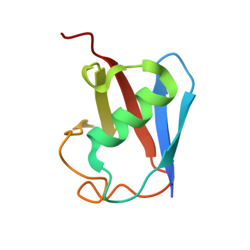
    5M93

  • PubMed Abstract: 

    Conventional ubiquitination involves the ATP-dependent formation of amide bonds between the ubiquitin C terminus and primary amines in substrate proteins. Recently, SdeA, an effector protein of pathogenic Legionella pneumophila, was shown to mediate NAD-dependent and ATP-independent ubiquitin transfer to host proteins. Here, we identify a phosphodiesterase domain in SdeA that efficiently catalyzes phosphoribosylation of ubiquitin on a specific arginine via an ADP-ribose-ubiquitin intermediate. SdeA also catalyzes a chemically and structurally distinct type of substrate ubiquitination by conjugating phosphoribosylated ubiquitin to serine residues of protein substrates via a phosphodiester bond. Furthermore, phosphoribosylation of ubiquitin prevents activation of E1 and E2 enzymes of the conventional ubiquitination cascade, thereby impairing numerous cellular processes including mitophagy, TNF signaling, and proteasomal degradation. We propose that phosphoribosylation of ubiquitin potently modulates ubiquitin functions in mammalian cells.


  • Organizational Affiliation

    Institute of Biochemistry II, School of Medicine, Goethe University Frankfurt, Theodor-Stern-Kai 7, 60590 Frankfurt am Main, Germany; Buchmann Institute for Molecular Life Sciences, Goethe University Frankfurt, Max-von-Laue-Str. 15, 60438 Frankfurt am Main, Germany.


Macromolecules
Find similar proteins by:  (by identity cutoff)  |  3D Structure
Entity ID: 1
MoleculeChains Sequence LengthOrganismDetailsImage
Polyubiquitin-B
A, B, C
80Homo sapiensMutation(s): 0 
Gene Names: UBB
UniProt & NIH Common Fund Data Resources
Find proteins for P0CG47 (Homo sapiens)
Explore P0CG47 
Go to UniProtKB:  P0CG47
PHAROS:  P0CG47
GTEx:  ENSG00000170315 
Entity Groups  
Sequence Clusters30% Identity50% Identity70% Identity90% Identity95% Identity100% Identity
UniProt GroupP0CG47
Sequence Annotations
Expand
  • Reference Sequence
Experimental Data & Validation

Experimental Data

  • Method: X-RAY DIFFRACTION
  • Resolution: 1.79 Å
  • R-Value Free: 0.231 
  • R-Value Work: 0.189 
  • R-Value Observed: 0.191 
  • Space Group: P 1 21 1
Unit Cell:
Length ( Å )Angle ( ˚ )
a = 31.596α = 90
b = 81.073β = 105.72
c = 51.287γ = 90
Software Package:
Software NamePurpose
REFMACrefinement
XDSdata reduction
SCALAdata scaling
PHASERphasing

Structure Validation

View Full Validation Report


Ligand Structure Quality Assessment 


Entry History & Funding Information

Deposition Data


Funding OrganizationLocationGrant Number
LOEWE program Ubiquitin Networks (Ub-NetGermanySPP 1580

Revision History  (Full details and data files)

  • Version 1.0: 2016-12-14
    Type: Initial release
  • Version 2.0: 2020-07-29
    Type: Remediation
    Reason: Carbohydrate remediation
    Changes: Atomic model, Data collection, Derived calculations, Structure summary
  • Version 2.1: 2024-01-17
    Changes: Data collection, Database references, Refinement description, Structure summary