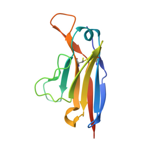
5L6Q

Refolded AL protein from cardiac amyloidosis


Experimental Data Snapshot

  • Method: X-RAY DIFFRACTION
  • Resolution: 1.40 Å
  • R-Value Free: 0.182 
  • R-Value Work: 0.147 
  • R-Value Observed: 0.149 

wwPDB Validation   3D Report Full Report


This is version 1.3 of the entry. See complete history


Literature

Common Fibril Structures Imply Systemically Conserved Protein Misfolding Pathways In Vivo.

Annamalai, K.Liberta, F.Vielberg, M.T.Close, W.Lilie, H.Guhrs, K.H.Schierhorn, A.Koehler, R.Schmidt, A.Haupt, C.Hegenbart, U.Schonland, S.Schmidt, M.Groll, M.Fandrich, M.

(2017) Angew Chem Int Ed Engl 56: 7510-7514

  • DOI: https://doi.org/10.1002/anie.201701761
  • Primary Citation of Related Structures:  
    5L6Q

  • PubMed Abstract: 

    Systemic amyloidosis is caused by the misfolding of a circulating amyloid precursor protein and the deposition of amyloid fibrils in multiple organs. Chemical and biophysical analysis of amyloid fibrils from human AL and murine AA amyloidosis reveal the same fibril morphologies in different tissues or organs of one patient or diseased animal. The observed structural similarities concerned the fibril morphology, the fibril protein primary and secondary structures, the presence of post-translational modifications and, in case of the AL fibrils, the partially folded characteristics of the polypeptide chain within the fibril. Our data imply for both analyzed forms of amyloidosis that the pathways of protein misfolding are systemically conserved; that is, they follow the same rules irrespective of where inside one body fibrils are formed or accumulated.


  • Organizational Affiliation

    Institute of Protein Biochemistry, Ulm University, Helmholtzstrasse 8/1, 89081, Ulm, Germany.


Macromolecules
Find similar proteins by:  (by identity cutoff)  |  3D Structure
Entity ID: 1
MoleculeChains Sequence LengthOrganismDetailsImage
H5AL
A, B
115Homo sapiensMutation(s): 0 
Entity Groups  
Sequence Clusters30% Identity50% Identity70% Identity90% Identity95% Identity100% Identity
Sequence Annotations
Expand
  • Reference Sequence
Small Molecules
Ligands 6 Unique
IDChains Name / Formula / InChI Key2D Diagram3D Interactions
PGE
Query on PGE

Download Ideal Coordinates CCD File 
R [auth B]TRIETHYLENE GLYCOL
C6 H14 O4
ZIBGPFATKBEMQZ-UHFFFAOYSA-N
PEG
Query on PEG

Download Ideal Coordinates CCD File 
I [auth A]DI(HYDROXYETHYL)ETHER
C4 H10 O3
MTHSVFCYNBDYFN-UHFFFAOYSA-N
ZN
Query on ZN

Download Ideal Coordinates CCD File 
C [auth A]
D [auth A]
E [auth A]
F [auth A]
J [auth B]
C [auth A],
D [auth A],
E [auth A],
F [auth A],
J [auth B],
K [auth B],
L [auth B]
ZINC ION
Zn
PTFCDOFLOPIGGS-UHFFFAOYSA-N
CO3
Query on CO3

Download Ideal Coordinates CCD File 
H [auth A],
P [auth B],
Q [auth B]
CARBONATE ION
C O3
BVKZGUZCCUSVTD-UHFFFAOYSA-L
CL
Query on CL

Download Ideal Coordinates CCD File 
O [auth B]CHLORIDE ION
Cl
VEXZGXHMUGYJMC-UHFFFAOYSA-M
NA
Query on NA

Download Ideal Coordinates CCD File 
G [auth A],
M [auth B],
N [auth B]
SODIUM ION
Na
FKNQFGJONOIPTF-UHFFFAOYSA-N
Experimental Data & Validation

Experimental Data

  • Method: X-RAY DIFFRACTION
  • Resolution: 1.40 Å
  • R-Value Free: 0.182 
  • R-Value Work: 0.147 
  • R-Value Observed: 0.149 
  • Space Group: P 21 21 2
Unit Cell:
Length ( Å )Angle ( ˚ )
a = 58.02α = 90
b = 105.81β = 90
c = 34.75γ = 90
Software Package:
Software NamePurpose
REFMACrefinement
XDSdata reduction
XSCALEdata scaling
PHASERphasing

Structure Validation

View Full Validation Report


Entry History 

Revision History  (Full details and data files)

  • Version 1.0: 2017-05-31
    Type: Initial release
  • Version 1.1: 2017-06-07
    Changes: Database references
  • Version 1.2: 2017-06-21
    Changes: Database references
  • Version 1.3: 2024-01-10
    Changes: Data collection, Database references, Derived calculations, Refinement description