5IUX

GLIC-V135C bimane labelled X-ray structure


Experimental Data Snapshot

  • Method: X-RAY DIFFRACTION
  • Resolution: 2.60 Å
  • R-Value Free: 0.237 
  • R-Value Work: 0.203 
  • R-Value Observed: 0.205 

wwPDB Validation   3D Report Full Report


Ligand Structure Quality Assessment 


This is version 1.3 of the entry. See complete history


Literature

Identification of a pre-active conformation of a pentameric channel receptor.

Menny, A.Lefebvre, S.N.Schmidpeter, P.A.Drege, E.Fourati, Z.Delarue, M.Edelstein, S.J.Nimigean, C.M.Joseph, D.Corringer, P.J.

(2017) Elife 6

  • DOI: https://doi.org/10.7554/eLife.23955
  • Primary Citation of Related Structures:  
    5IUX

  • PubMed Abstract: 

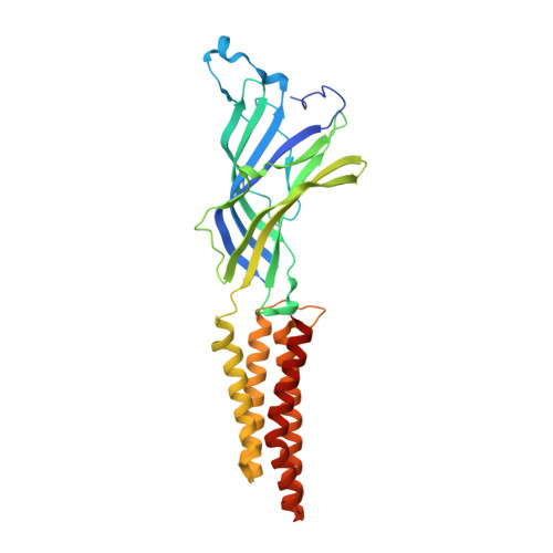
    Pentameric ligand-gated ion channels (pLGICs) mediate fast chemical signaling through global allosteric transitions. Despite the existence of several high-resolution structures of pLGICs, their dynamical properties remain elusive. Using the proton-gated channel GLIC, we engineered multiple fluorescent reporters, each incorporating a bimane and a tryptophan/tyrosine, whose close distance causes fluorescence quenching. We show that proton application causes a global compaction of the extracellular subunit interface, coupled to an outward motion of the M2-M3 loop near the channel gate. These movements are highly similar in lipid vesicles and detergent micelles. These reorganizations are essentially completed within 2 ms and occur without channel opening at low proton concentration, indicating that they report a pre-active intermediate state in the transition pathway toward activation. This provides a template to investigate the gating of eukaryotic neurotransmitter receptors, for which intermediate states also participate in activation.


  • Organizational Affiliation

    Channel Receptors Unit, Institut Pasteur, Paris, France.


Macromolecules
Find similar proteins by:  (by identity cutoff)  |  3D Structure
Entity ID: 1
MoleculeChains Sequence LengthOrganismDetailsImage
Proton-gated ion channel
A, B, C, D, E
317Gloeobacter violaceus PCC 7421Mutation(s): 1 
Gene Names: glvIglr4197
Membrane Entity: Yes 
UniProt
Find proteins for Q7NDN8 (Gloeobacter violaceus (strain ATCC 29082 / PCC 7421))
Explore Q7NDN8 
Go to UniProtKB:  Q7NDN8
Entity Groups  
Sequence Clusters30% Identity50% Identity70% Identity90% Identity95% Identity100% Identity
UniProt GroupQ7NDN8
Sequence Annotations
Expand
  • Reference Sequence
Small Molecules
Ligands 6 Unique
IDChains Name / Formula / InChI Key2D Diagram3D Interactions
PC1
Query on PC1

Download Ideal Coordinates CCD File 
EA [auth D]
FA [auth D]
G [auth A]
GA [auth D]
H [auth A]
EA [auth D],
FA [auth D],
G [auth A],
GA [auth D],
H [auth A],
I [auth A],
P [auth B],
PA [auth E],
QA [auth E],
R [auth B],
RA [auth E],
S [auth B],
W [auth C],
Y [auth C],
Z [auth C]
1,2-DIACYL-SN-GLYCERO-3-PHOSPHOCHOLINE
C44 H88 N O8 P
NRJAVPSFFCBXDT-HUESYALOSA-N
LMT
Query on LMT

Download Ideal Coordinates CCD File 
F [auth A]
K [auth A]
NA [auth E]
OA [auth E]
Q [auth B]
F [auth A],
K [auth A],
NA [auth E],
OA [auth E],
Q [auth B],
X [auth C]
DODECYL-BETA-D-MALTOSIDE
C24 H46 O11
NLEBIOOXCVAHBD-QKMCSOCLSA-N
6E3
Query on 6E3

Download Ideal Coordinates CCD File 
HA [auth D],
L [auth A]
2,3,5,6-tetramethyl-1H,7H-pyrazolo[1,2-a]pyrazole-1,7-dione
C10 H12 N2 O2
VWKNUUOGGLNRNZ-UHFFFAOYSA-N
BR
Query on BR

Download Ideal Coordinates CCD File 
AA [auth C],
IA [auth D],
M [auth A],
SA [auth E],
T [auth B]
BROMIDE ION
Br
CPELXLSAUQHCOX-UHFFFAOYSA-M
ACT
Query on ACT

Download Ideal Coordinates CCD File 
CA [auth C]
DA [auth C]
J [auth A]
KA [auth D]
LA [auth D]
CA [auth C],
DA [auth C],
J [auth A],
KA [auth D],
LA [auth D],
MA [auth E],
N [auth A],
O [auth A],
UA [auth E],
V [auth B]
ACETATE ION
C2 H3 O2
QTBSBXVTEAMEQO-UHFFFAOYSA-M
NA
Query on NA

Download Ideal Coordinates CCD File 
BA [auth C],
JA [auth D],
TA [auth E],
U [auth B]
SODIUM ION
Na
FKNQFGJONOIPTF-UHFFFAOYSA-N
Experimental Data & Validation

Experimental Data

  • Method: X-RAY DIFFRACTION
  • Resolution: 2.60 Å
  • R-Value Free: 0.237 
  • R-Value Work: 0.203 
  • R-Value Observed: 0.205 
  • Space Group: C 1 2 1
Unit Cell:
Length ( Å )Angle ( ˚ )
a = 182.034α = 90
b = 134.075β = 102.51
c = 159.945γ = 90
Software Package:
Software NamePurpose
BUSTERrefinement
XDSdata reduction
SCALAdata scaling
REFMACphasing

Structure Validation

View Full Validation Report


Ligand Structure Quality Assessment 


Entry History 

Deposition Data

Revision History  (Full details and data files)

  • Version 1.0: 2017-03-29
    Type: Initial release
  • Version 1.1: 2018-01-31
    Changes: Database references
  • Version 1.2: 2018-05-09
    Changes: Data collection, Database references
  • Version 1.3: 2019-10-16
    Changes: Data collection